Request a video call
Confirm Your Virtual Consultation
Prostate Cancer Center Profile Overview

Specialty Prostate Cancer Center
The Prostate-Center is part of the Prof. Dr. Stehling Institute for Diagostic Imaging. It was founded in 2010 by Prof. Michael K. Stehling.
 At the Prostate-Center physicians, scientists and engineers collaborate closely.
cancer One goal is the further development of minimally-invasive diagnostic techniques and therapies for diseases of the prostate and to make them available for the treatment of patients.
Prostate cancer (PCa) constitutes one of the greatest challenges in medical care of men.
With 25 % of all cancers, prostate cancer is the most frequent malignant tumour in men and responsible for 10 % of all deaths. Currently established treatment methods for prostate cancer are invasive an in many cases impair the quality of life of the afflicted men through impotence and incontinence.
At the same time, many prostate cancers do not have to be treated at all, because they remain confined to the prostate gland, causing neither symptoms nor leading to death.

At the Prostate-Center we thus employ the most advanced methods for the diagnosis and therapy of prostate cancer. In our effort, two goals are paramount: Survival and quality of life.
To achieve this, we are have established a close interdisciplinary collaboration with Urologists, Pathologists, Radiologist, Anaesthesiologists and Scientists from different countries in Europe, USA and Asia. Continuous innovation and the highest possible degree of competence are thus guaranteed.
Phototherapy
Light is not only a natural part of our environment, it also plays an important role in maintaining one's health and well-being. With this in mind, our interior design team created a open space with this objective: out with the sterile hospital 'look' and drab treatment rooms; in with a modern, relaxed and comfortable environment.

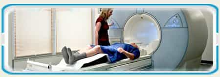
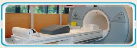
Neutral colors, discreet lighting and simple design invite our patients to spend a stress-free and comfortable time with us. These elements are used throughout the practice including in our treatment rooms: our specially designed MRI examinations rooms provide patients with natural lighting and an outside view of the scenery outside our offices.
Our Physicians

The team of diagnostic imaging experts with years of experience in treating their fields after the state of the science. They are guided by international standards of diagnosis and treatment as well as to the guidelines of leading hospitals, medical societies and government institutions.
Prof. Dr. Phil. Dr. Med. Michael K. Stehling
Director of the Institute, Head of Research and Development
Dr. Stephen Zapf
Deputy Director, Head of cross-sectional imaging and micro-therapy.
Prostate Cancer: Radical Treatments Bring Irreversible Side Effects
One out of 6 men develops prostate cancer. Contrary to the benign enlargement of the prostate (BPH) prostate cancer does not cause any symptoms.
Studies have represented also, that:
-
New cases of prostate cancer appear every 2.4 minutes.
-
Non-smoking men are more likely to develop prostate cancer than other cancer forms, like colon, bladder, melanoma, lymphoma and kidney cancer.
-
Probability to be diagnosed with prostate cancer by the men is 35% more than by the women to be diagnosed with breast cancer.
-
Small prostate cancer can start developing during a man’s 30s, and the appearance rate increases by around 10% every 10 years of life (50% in 50s, 60% in 60s, etc.).
-
Nearly 88 men only in USA die from it every day
Despite being frequent, many prostate cancers are relatively innocuous: Many men carry undetected prostate cancers inside their bodies which will remain asymptomatic and will not shorten their lifespan.
Unfortunately men are not always comprehensively informed about the severe “collateral damage” of this treatment which is appropriately called “radical”: After prostatectomy 70 % of all men are impotent (not able to have an erection anymore) and 10 – 50 % incontinent (not able hold their urine).
Recommendations
Don’t panic when the diagnosis of prostate cancer is first established: In most cases the cancer has been inside the body for many years already. It grows very slowly. You have plenty of time to collect information about treatment methods.
A precise diagnostic work-up is of paramount importance for the choice of the optimal treatment regime.
Our Treatments
-
Prostate Cancer: Screening and Diagnosis
-
Multi-modal MR-Diagnostic Imaging
-
Prostate Cancer Treatments
-
3D-Biopsy
-
Treatment of Prostate Cancer with Irreversible Electroporation (NanoKnife)
New Benchmark for Prostate Cancer Treatment
New treatment method for prostate cancer – NanoKnife (irreversible electroporation or IRE), affords new possibilities for focal prostate therapy with only minimal side effects and sets the new benchmark in this field.
Whilst currently established treatment methods cause impotence (loss of erectile function of the penis) and incontinence (loss of urinary bladder control) in the majority of men, these side effects can be avoided in most cases with irreversible electroporation.
The significance of paradigm change in prostate therapy becomes clear when one considers that every 6th man is affected by prostate cancer. And whilst prostate cancer was primarily a disease of elderly men in the past, today younger, too, are increasingly afflicted.
Nanoknife-Treatment
 Treatment with irreversible electroporation (IRE), the NanoKnife, causes cell death induced by strong local electric fields. This, however, only affects parenchymal and tumor cells.
Nerves and vessels are spared or only partially damaged. This was the neurovascular bundle, responsible for penile erection and urinary continence, remains intact.
Modern Treatment Methods
With this method the novel, gentle and minimally-invasive treatment method has become available for patients at the Prostate-Center, method which affords a therapy of prostate cancer without grave side effects.
Do you need treatment for Prostate Cancer?
The golden rule of medicine to increase your chances of a recovery is to identify the risk and treat the disease as early as it possible.
In order to identify the risk of the prostate cancer for you special diagnostic work-up is necessary.
Prostate Cancer Center, Frankfurt, Germany Profile Details
|
About Prostate Cancer Center
The Prostate-Center is part of the Prof. Dr. Stehling Institute for Diagostic Imaging. It was founded in 2010 by Prof. Michael K. Stehling. At the Prostate-Center physicians, scientists and engineers collaborate closely. One goal is the further development of minimally-invasive diagnostic techniques and therapies for diseases of the prostate and to make them available for the treatment of patients.
Currently established treatment methods for prostate cancer are invasive an in many cases impair the quality of life of the afflicted men through impotence and incontinence. At the same time, many prostate cancers do not have to be treated at all, because they remain confined to the prostate gland, causing neither symptoms nor leading to death. A paradigm change in the treatment of prostate cancer is thus necessary: Instead of a radical therapy with massive side effects a minimally-invasive therapy without or with only minimal side effects should be employed. As long as the spread of the cancer in the body is prevented, the therapy is successful. At the Prostate-Center we thus employ the most advanced methods for the diagnosis and therapy of prostate cancer. In our effort, two goals are paramount: Survival and quality of life. To achieve this, we are have established a close interdisciplinary collaboration with Urologists, Pathologists, Radiologist, Anaesthesiologists and Scientists from different countries in Europe, USA and Asia. Continuous innovation and the highest possible degree of competence are thus guaranteed.
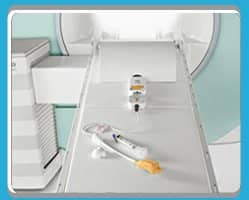
Neutral colors, discreet lighting and simple design invite all our patients to spend a stress-free and comfortable time with us. These are the elements used throughout the practice including in our treatment rooms: our specially designed MRI examinations rooms provide patients with natural lighting and an outside view of the scenery outside our offices. Our open MRI machine and large window help to avoid the feeling of suffocation or claustrophobia. Come and see yourself!
Call us now to schedule an appointment and meet our team of trained experts in a calm and relaxing atmosphere as you learn about and consider the best treatment options that are right for you and your individual needs and concerns.
|
Prostate Cancer Center Treatments Offered
|
Our Treatments
Which treatment is chosen for which patient depends on many factors: Besides the tumor stage and grade the choice is also influenced by the patient’s age and health, but also by personal preferences. ? When the carcinoma is confined to the prostate, a focal treatment method which focuses on the prostate can be chosen. The current routine focal treatment method is the surgical removal of the prostate (radical prostatectomy), also with robotically-assisted surgery (so-called Da Vinci Surgery). Other treatment methods comprise brachytherapy (internal radiation treatment through implanted radioactive seeds) or external radiation therapy. All these radical methods, however, have severe side effects and result in erectile impotence in the majority of patients and cause urinary incontinence in up to 50%. Thermal methods like focused ultrasound (HIFU) cryotherapy still have vast side effects and frequent reoccurrences. When the cancer has spread beyond the capsule, hormone therapy (blockade of male sexual hormones) or external radiation therapy constitute the primary choices for treatment. Such treatments have also adverse side effects.
Important structures of the prostate such as vessels and nerves, the base of the bladder, the urinary sphincter and the bowel, remain intact. Contrary to other treatment methods impotence and incontinence can thus be avoided, likewise wound pain and scar formation. Treatment is complete within 24 hours. Rehabilitation and after-care are not necessary. In advanced cases of prostate cancer, the NanoKnife treatment can be complemented with a cyberknife treatment.
What is Prostate Cancer?
Prostate cancer occurs when cells in the prostate gland grow out of control and the ability to regulate cell growth or death is lost. The enlargement of extra cells forms a mass of tissue called a growth or tumor. Actually, it is one of the most common cancer forms, affecting 1 in 6 men. Prostate growths can be divided into two groups: Benign (not cancer), such as Benign prostatic hyperplasia (BPH), enlarged prostate or prostatitis, which according to a common belief may lead over time to the development of prostate cancer. And Malignant (cancer) tumors. As opposed to benign it:
Prostate Cancer Symptoms
Some patients can have an experience with changes in urinary or sexual function which might indicate the presence of prostate cancer. These symptoms include, for example:
Early Detection and Prevention of
Prostate Cancer
The early detection of the cancer cells plays here the utmost important role. Screening for prostate cancer usually comprises blood testing for PSA (prostate-specific antigen) and a digital rectal exam of the prostate gland. Whilst easy to perform and cheap, prostate cancer can be missed by these tests. On the other hand “false positive” findings can mimick cancer, albeit being benign changes of no importance. Thanks to new developments in this filed, the identification of the precancerous prostate tumors can be found in much earlier stages and hence leaving the patient more gentle treatment options and a higher chance of survival. Multiparametric MRI-Scans, Cholin-PET scans, MRI-Guided 3d-Biopsies are advanced tools giving rise to a new era of prostate cancer treatment.
Prostate Cancer: Screening and Diagnosis
When I should make a screening?
Therefore, annual prostate cancer screening is recommended for men aged 50 or above. For patients who have first degree relatives with prostate cancer, screening should commence at the age of 40. Routine screening is not accurate enough Screening for prostate cancer usually comprises blood testing for PSA (prostate-specific antigen) and a digital rectal exam of the prostate gland. Whilst easy to perform and cheap, prostate cancer can be missed by these tests. On the other hand “false positive” findings can mimic cancer, albeit being benign changes of no importance. Percentage of men harboring a prostate cancer (PC) as a function of the PSA-level. Whilst in Germany a biopsy is recommended with PSA > 4 ng/ml, in the US the threshold for biopsy is 2.5 ng/ml. Ordinary biopsy defines the diagnosis only in up to 35% of cases. MRI-Guided 3d-Biopsies are much more precise, have a much lower infection rate and are absolutely necessary for every modern focal treatment.
“Biopsy-Free” Diagnostic of the Prostate
More Accurate and Safe
As an alternative to a biopsy a non-invasive (“biopsy-free”) diagnostic work-up can be chosen initially, employing endorectal magnetic resonance imaging (MRI) and spectroscopy (MSR). Advantages of the “Biopsy-Free”-Diagnostic:
Multi-parametric MR Imaging Diagnostic
Most Accurate Imaging Technique for Prostate Cancer Detection
Due to this, MRI is superior to other imaging techniques such as ultrasound and also when compared to elastography. The four pillars of multi-modal MRI diagnostics of prostate cancer: Imaging, diffusion, perfusion and spectroscopy. Also the analysis of the chemical composition of prostatic tissue makes the diagnostic with MSR (MR-Spectroscopy) so precise and valuable. Prostate spectroscopy depicts the chemical make-up of prostatic cells. Healthy cells contain large amounts of citrate and little cholin. Cancer cells contain less citrate, which is metabolised for the generation of energy, and more cholin, which is needed for the assembly of the cell membranes in the tumour cells.
Our Treatment of Prostate Cancer
Precise and Safe
Step 1: Diagnostics
Specifying the Target with Transperineal 3D-Biopsy of the Prostate: Pain-free – Safe – Precise Pain-Free and Safe Transperineal biopsy is carried out pain-free in full anaesthesia, and steril under operation room conditions such that the risk of a post biopsy infection of the prostate (prostatitis) is negligible. Precise
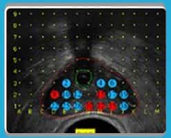
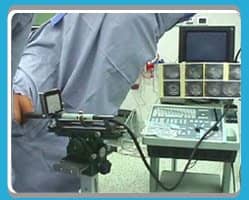
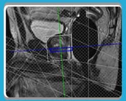
A 3D biopsy is carried out ultrasound-guided with the aid of a “stepper” and “grid”. The MRI data give additional insight and allow a precise planning. Also the results of the biopsy can be displayed within the MRI pictures so that a very precise treatment planning is possible. This particular method makes it possible to sample tissue from the whole prostate in well-defined locations. This way, a three-dimensional tissue map can be assembled, which reveals whether a prostate cancer is confined to a specific location in the prostate (focal) or distributed over an extended volume (multi-focal). In advanced cases a whole body MRI determines the stage of the cancer by looking for possible metastasis. Advantages of the 3D-Biopsy: 3D-Biopsy vs. usual Biopsy
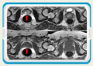
MRI scans of the prostate before (left) and after (right) 3D-biopsy with an average of 60 samples.
Step 2: Therapy with the NanoKnife and the CyberKnife
?Non-cellular tissue components are not damaged by the NanoKnife-Treatment. The tissue matrix, which is made up of collagen and elastin fibres, proteoglycans, etc., remains fully preserved. Because blood and lymphatic vessels as well as the urethra, besides cells, have an extensive and stable tissue matrix, they regenerate fully: The destroyed cells re-colonise along the matrix. ?Other treatment methods, which destroy all tissue elements, induce a tissue necrosis (coagulation of all tissue components). This necrosis is eventually removed over the course of many months by an inflammatory process encompassing the necrosis. This process causes inflammatory pain and, after healing of the necrosis, scarring, which can severely hamper subsequent treatments. ?NanoKnife or Irreversible Electroporation, on the contrary, does not induce any tissue necrosis and consequently no inflammation. Treatment with the NanoKnife thus avoids pain and scarring. Because blood vessels and tissue perfusion are preserved the dead cell’s debris is removed quickly from the treatment zone. If the prostate cancer has spread, the metastasis can an many cases be treated with a Cyberknife system. The Cyberknife system is a computer operated robot - guided radiation therapy approach which allows a very precise radiation deployment, even more precise than proton therapy.
Outline of the 3D-Biopsy-Work-Up and Nanoknife-Treatment
Step 1: Preparation for the Treatment -3D-Biopsy-Work-Up
1 Day: Localisation of the tumor cells with 3D-Biopsy-Work-Up?2 Day: Histological investigation and MRI-Control Step 2: Nanoknife Treatment
3 Day: Prostate Cancer Nanoknife Treatment?4 Day: MRI-Control and Discharge from the hospital
|
Prostate Cancer Center Certificates, Accreditations, Qualifications Treatments Offered
|
Experience and Commitment
In the center of the Rhine-Main area, the diagnostic imaging in alpha-house in May 2005 phil under the direction of Prof. Dr. Dr.. Dr. med. Michael K. Stehling continued at a new site. Following on from his experience at Harvard Medical School and the long-standing collaboration with the Nobel Prize in Medicine in 2003, Sir Peter Mansfield sets, Prof. Stehling here continues its commitment to health care and a precise and gentle early detection of diseases.
The team of diagnostic imaging experts with years of experience in treating their fields after the state of the science. They are guided by international standards of diagnosis and treatment as well as to the guidelines of leading hospitals, medical societies and government institutions.
Far Ahead to Knowledge and Technology
For you as a patient, this will require high medical expertise and use of the latest high-tech methods. Personal service and friendly atmosphere complete our performance for you. Our tight integration with a network of specialists also your follow-up is also on the investigation into the diagnostic imaging in good hands.
Our Physicians
Prof. Dr. phil. Dr. med. Michael K. Stehling
After completing his studies in Human Sciences and Medical Physics at the University of Manchester, England and the Johann-Wolfgang-Goethe University in Frankfurt, Germany Dr. Stehling served as Research Assistant to Sir Peter Mansfield, Prof of Physics and Nobel Prize winner in Medicine in 2003 (for discoveries concerning magnetic resonance imaging) and thereafter completed doctorate work in echo-planar imaging. He completed his radiology training at the University Hospital in Zurich, Switzerland and was later Resident and Clinical Fellow in Radiology at the Beth Israel Hospital at Harvard Medical School in Boston, MA, USA. During this time he also worked with Siemens AG in Erlangen in their Research and Development department. Prior to founding and starting the Institut fur Bildgebende Diagnostik in Frankfurt/Offenbach in 2005, Dr. Stehling served as Senior Physician and Director of the Radiology department in the Radiology Institute at the Ludwig-Maximilians Unversities, Grosshadern Clinic. During this time he completed his qualification as an Adjunct Professor and gave regular lectures at LMU. Since 2009 he has been Associate Professor of Radiology at the Boston Univeristy School of Medicince, Department of Radiology. He is a member of the German Radiological Society, the Radiological Society of North America and the International Society for Magnetic Resonance in Medicine.
Dr. Stephen Zapf
Dr. Kirsten Holsteg
Dr. Cristian Rodriguez
|
Prostate Cancer Center Testimonials
Prostate Cancer Center Awards & Recognitions
Frankfurt, Germany Destination Overview
Location

About Medical Center
- Speciality: Cancer Treatment, ENT, Executive Healthcheck, Gynecology Treatment, Organ Transplant, Urology
- Location: Strahlenberger Straße 110 63067 , Frankfurt, Germany
- Medical Center Prices: Prostate-Cancer-Center
- Overview: Prostate Cancer Center is the best medical center for the Prostate Treatments and Diagnosis in Europe. Best treatment for prostate cancer using most efficient technologies such as nanoknife, MRI and 3D Biopsy. Find and compare your treatment options here.



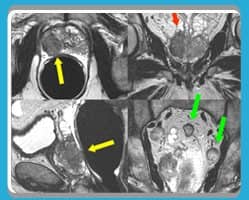

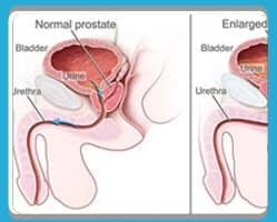


 Prostate cancer (PCa) constitutes one of the greatest challenges in medical care of men. With 25 % of all cancers, prostate cancer is the most frequent malignant tumour in men and responsible for 10 % of all cancer deaths.

 Prostate cancer is a form of cancer that develops in the prostate (gland of the male reproductive system).
 Not every patient has symptoms of prostate cancer and the first signs of prostate cancer are first detected by a doctor during the check-up or diagnostic.
 As every untreated and allowed to grow, malignant cells from cancerous tumors can spread and cause metastasis in other part of the body. If it happens, the chances to treat the disease successfully much lower.
 Prostate cancer occurs with increasing frequency, even in younger men. Contrary to benign prostatic hypertrophy (BPH) or prostatitis most prostate cancers do not cause any symptoms and are visible when it is too late.
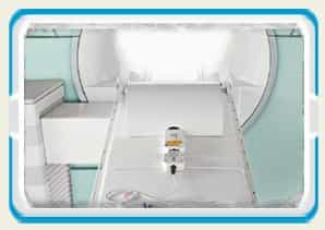 Contrary to other imaging techniques (Computed tomography, ultrasound imaging, szintigraphy, positron emission tomography) MRI provides multiple mutually independent parameters for the assessment of the prostate:
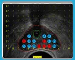 The highest accuracy is achieved with transperineal (through the skin of the pelvic floor) 3D-biopsy, which affords a detection rate for prostate cancer between 75 and 100 %.
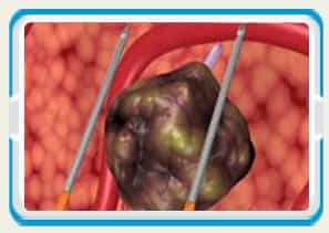 Nanoknife is a novel promising method for the selective destruction of tumor cells by strong local electric fields. Affected by the electric fields of several thousand volts cell membranes are opened up until the cell ruptures. This process corresponds to an induced apoptosis (natural cell death).




Share this listing