
What Is ESD Endoscopic Resection in Gangnam?
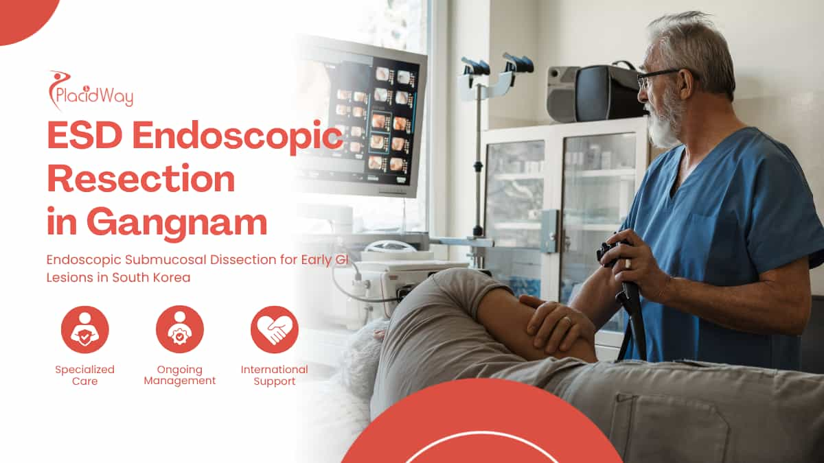
The journey toward Endoscopic Submucosal Dissection (ESD) rarely begins with the intention to seek complex treatment. It typically starts with a routine screening, a follow-up endoscopy for persistent indigestion, or a colonoscopy designed to prevent problems rather than find them. When the physician calls to discuss the biopsy results, the initial diagnosis of an early gastrointestinal lesion or high-grade dysplasia often brings a confusing mix of immense relief and sudden clinical tension.
When screening reveals an early gastrointestinal lesion or high-grade dysplasia, patients may face difficult treatment decisions. For selected cases, ESD endoscopic resection in Gangnam offers a minimally invasive option for removing early GI lesions.
The Decision Fork: Efficacy Versus Organ Preservation
Patients often find themselves caught between two highly undesirable extremes. They can opt for standard Endoscopic Mucosal Resection (EMR), which might require taking the lesion out in multiple fragments and risking recurrence, or they can choose open or laparoscopic surgery, which guarantees the removal of the disease but permanently alters their digestive anatomy. ESD represents a highly technical middle path—allowing the entire lesion to be peeled away from the inside without removing the organ itself.
Recognizing that ESD exists is only the first step in the patient's research process. The subsequent realization is that this procedure demands a high level of technical skill, a steep learning curve, and an environment characterized by high procedural volume.
This realization frequently prompts patients to look beyond their local healthcare networks and consider specialized centers in destinations like Gangnam, Seoul, where unique national health policies have transformed a rare intervention into a daily clinical routine.
Anatomy of the Dissection Plane: Why Depth Changes Everything
To understand why a seemingly simple superficial lesion requires such a specialized intervention, it is necessary to visualize the structure of the gastrointestinal tract. The wall of the stomach, esophagus, or colon is not a single solid barrier. It is constructed from distinct biological layers, much like heavily layered wallpaper applied over a muscular foundation.
When early cancers or precancerous dysplasias begin to grow, they almost exclusively start on the innermost surface. The medical challenge lies in determining exactly how deeply these abnormal cells have rooted themselves into the underlying tissue. The entire justification for endoscopic intervention relies on the premise that the disease has not yet reached the deeper biological highways where cancer cells can travel to other parts of the body.
- The Mucosa: This is the innermost lining that interacts directly with food and digestive acids. Early lesions originate here, and if they remain strictly confined to this topmost layer, the risk of lymph node metastasis is generally extremely low.
- The Submucosa: Situated just beneath the mucosa, this spongy connective tissue serves as the critical workspace for the endoscopist during ESD. It contains a rich network of blood vessels and lymphatic channels. If a tumor invades deeply into this layer, the risk of spread increases, potentially changing the treatment from endoscopy to surgery.
- The Muscularis Propria: This thick muscle layer is responsible for the contractions that move food through the digestive system. If an endoscopic instrument cuts too deeply and breaches this muscle layer, a perforation occurs, which is a serious complication that requires immediate closure or surgical repair.
Traditional snare techniques often struggle with lesions that are flat, laterally spreading, or larger than two centimeters because the wire loop cannot effectively grab a wide, shallow area of tissue. If the physician attempts to snare a large flat lesion, they are forced to remove it in multiple pieces. This destroys the edges of the tissue sample, leaving pathologists unable to confirm whether the margins are completely clear of abnormal cells.
The Three Clinical Paths: EMR, Organ Surgery, and ESD
When consulting with a gastroenterologist or a surgical oncologist, the patient is typically evaluating a spectrum of invasiveness. Understanding the mechanical differences between these three primary approaches is essential for setting realistic expectations and navigating the trade-offs of each.
The distinction between these methods often hinges on the concept of en bloc resection. This term simply means removing the targeted tissue in one single, unbroken piece, rather than tearing it into fragments. A perfectly intact specimen allows the laboratory to definitively map the boundaries of the disease, which is the ultimate goal of any oncological or preventative resection.
| Endoscopic Mucosal Resection (EMR) | Endoscopic Submucosal Dissection (ESD) | Surgical Resection |
|---|---|---|
| Mechanism: Fluid is injected under the lesion to lift it, and a wire snare is looped over the top to slice it off. | Mechanism: A specialized electrosurgical knife dissects the submucosal layer beneath the lesion, peeling it away completely. | Mechanism: Laparoscopic or open surgery removes the affected section of the organ, along with surrounding lymph nodes. |
| Best For: Small (under 2cm), raised lesions that can easily fit inside a single snare loop. | Best For: Large, flat, or laterally spreading lesions that must be removed in one piece (en bloc). | Best For: Cancers that have invaded deeply into the submucosa or present a high risk of lymph node involvement. |
| Limitations: Large lesions require piecemeal removal, increasing the risk of local recurrence and complicating pathology. | Limitations: Highly technically demanding, requires a long procedure time, and carries risks of bleeding and perforation. | Limitations: Major physical trauma, permanent anatomical changes, and longer, more painful recovery periods. |
For a patient who has been told they need a partial gastrectomy for an early, flat mucosal cancer, ESD offers a profound alternative. It provides the oncological certainty of a complete margin assessment without the life-altering consequences of losing a portion of the digestive tract. However, identifying a hospital with a team capable of performing this delicate peeling process safely is a significant hurdle.
The Impact of National Screening on South Korean Expertise
The geographical element of a patient's search often shifts to East Asia due to distinct epidemiological realities. South Korea has historically experienced some of the highest incidence rates of gastric cancer in the world. In response, the South Korean government implemented a National Cancer Screening Program in the early 2000s, which provides regular screening endoscopies for citizens over the age of forty.
This nationwide policy fundamentally altered the landscape of gastrointestinal medicine in the country. By screening asymptomatic adults, physicians began detecting thousands of gastric lesions at incredibly early, asymptomatic stages. If surgeons had removed the stomachs of every patient diagnosed with these early lesions, the national morbidity rate would have been catastrophic. Instead, South Korean endoscopists became highly incentivized to perfect and scale minimally invasive endoscopic resections.
The Volume-Expertise Connection
In medical procedures characterized by a steep learning curve, institutional volume is a primary driver of safety and success. While a specialist in a Western hospital might perform a few dozen ESD procedures a year, specialized endoscopists in Gangnam's tertiary hospitals often perform hundreds. This immense repetition refines the micro-movements required to dissect tissue millimeters away from the organ's muscle wall, directly impacting the rates of complete resection and complication management.
For an international patient traveling to Seoul, accessing clinics in Gangnam is not merely about seeking out a medical tourism hub; it is about plugging into a healthcare infrastructure where this specific, highly demanding procedure is treated as a routine daily occurrence. The concentration of university-affiliated hospitals and specialized GI centers in this district means that the entire clinical team—from the primary endoscopist to the assisting nurses and the evaluating pathologists—is acutely familiar with the nuances of submucosal dissection.
Inside the Submucosal Space: How the Procedure Unfolds
Understanding what actually happens while the patient is sedated helps demystify the procedure and clarifies why institutional expertise is so vital. ESD is essentially a microscopic surgery performed through a long, flexible tube, relying entirely on the visual feed from a high-definition camera and the manipulation of millimeter-scale instruments.
The intervention requires significant patience and precision. Unlike a standard EMR, which might take fifteen minutes, an ESD procedure can take anywhere from one to several hours, depending on the size and location of the lesion. The treating physician must navigate a highly vascular environment while constantly adjusting the endoscope to maintain a clear line of sight.
The Sequential Steps of Dissection
- Mapping and Marking: Using advanced imaging techniques, such as Narrow Band Imaging (NBI) or specialized dyes (chromoendoscopy), the physician clearly identifies the exact boundaries of the dysplastic tissue. Tiny burn marks are made around the perimeter to guide the cutting path.
- Submucosal Injection: A viscous fluid, often containing hyaluronic acid mixed with a blue dye and adrenaline, is injected directly beneath the lesion. This fluid acts as a hydraulic cushion, physically lifting the diseased mucosa away from the dangerous underlying muscle layer.
- Circumferential Incision: Using an electrosurgical knife, the endoscopist carefully cuts along the previously marked dots, creating a full circle around the lesion and breaching the mucosal surface to expose the blue-tinted submucosal cushion beneath.
- Submucosal Dissection: This is the core of the procedure. The physician uses the electrosurgical knife to carefully tease apart and cut the connective tissue fibers within the submucosal space, effectively tunneling beneath the lesion until it is completely freed from the organ wall.
- Hemostasis and Closure: Throughout the dissection, exposed blood vessels are carefully coagulated using thermal forceps to prevent bleeding. Once the tissue is removed, the remaining artificial ulcer is inspected, and any visible vessels are sealed. In some cases, metallic clips are applied to close the defect or prevent delayed hemorrhage.
Organ-Specific Microenvironments: Stomach, Colon, and Esophagus
While the fundamental principles of ESD remain consistent across the digestive tract, the physical realities of the stomach, colon, and esophagus create entirely different technical challenges. A patient researching a gastric lesion is facing a very different risk profile compared to a patient dealing with a colonic tumor. The environment dictates the complexity of the dissection.
Gastric (Stomach) ESD
The stomach is where ESD was originally pioneered. It provides a relatively spacious cavity, allowing the endoscope more room to maneuver and retroflex (bend backward). However, the gastric wall has a rich, complex blood supply. The primary challenge here is managing intraoperative and delayed bleeding, requiring the endoscopist to identify and meticulously seal numerous submucosal vessels during the dissection process.
Colorectal (Colon & Rectum) ESD
The colon represents a much more hostile environment for endoscopic surgery. The colonic wall is exceptionally thin, often measuring just a few millimeters, making the margin for error incredibly small. The primary fear during colorectal ESD is perforation. Additionally, the colon is a winding, tubular structure that constantly moves with respiration and peristalsis, making stabilization of the endoscope a major technical hurdle.
Esophageal ESD
The esophagus is a narrow, straight tube that leaves very little room for instrument angulation. While perforation remains a risk, the most significant long-term concern with esophageal ESD is stricture formation. If a large lesion is removed that encircles more than three-quarters of the esophageal circumference, the resulting scar tissue can contract as it heals, physically narrowing the throat and causing difficulty swallowing.
Recognizing these anatomical realities emphasizes why pre-procedural imaging and expert assessment are critical. The treating team in South Korea will thoroughly review the lesion's characteristics—such as size, location, and lifting signs—to ensure that the chosen technique aligns safely with the specific organ's limitations.
The Pathology Report: The True Moment of Continuity
A common misunderstanding among patients is that the tension resolves the moment they wake up in the recovery room and are told the procedure went well. In reality, a successful visual removal in the endoscopy suite is only provisional. The definitive outcome is entirely dependent on the microscopic evaluation of the tissue that was removed.
Once the lesion is extracted en bloc, it is carefully pinned to a flat board, preserved, and sent to the pathology laboratory. The pathologist sections the tissue into tiny slices to evaluate the depth of cellular invasion and to check the lateral (side) and vertical (deep) margins. This evaluation determines whether the resection was curative or non-curative, representing the most pivotal fork in the patient's long-term care plan.
Curative vs. Non-Curative Resection
- Curative Resection: The pathology report confirms that the margins are entirely clear of disease, the cancer cells did not invade deeply into the submucosal layer, and there is no evidence of cancer in the microscopic blood or lymphatic vessels (no lymphovascular invasion). In this scenario, the ESD is considered a complete success, and the patient transitions to routine surveillance.
- Non-Curative Resection: The report reveals that the tumor had invaded deeper than pre-operative estimates suggested, or that malignant cells are present at the deep margin or within the lymphatic channels. If this occurs, the risk of residual cancer or lymph node metastasis is deemed too high. The patient will likely be advised to undergo the very surgical resection (gastrectomy or colectomy) they initially hoped to avoid.
This uncertainty must be managed emotionally before traveling. ESD is both a therapeutic intervention and the most accurate diagnostic biopsy possible. Patients must understand that while the intent is curative, the biology of the tumor dictates the final result. If the pathology necessitates further surgery, the ESD was not a failure; it provided the precise staging information required to save the patient's life.
Risks, Complications, and Recovery Reality
Because ESD is a highly specialized, tissue-altering procedure, it carries specific risks that must be weighed carefully against the benefits of organ preservation. The artificial ulcer left behind inside the digestive tract takes several weeks to heal. During this time, the patient must remain vigilant for delayed complications, which can present days after the initial intervention.
Monitoring for Post-Procedure Complications
Patients are closely observed for symptoms that require immediate medical attention during the initial healing phase. Standard protocols prioritize identifying two primary events:
- Delayed Bleeding: Even when visible vessels are meticulously sealed, digestive acids or mechanical stress can cause a vessel to open later. This is indicated by black, tarry stools, vomiting blood, dizziness, or sudden extreme weakness.
- Delayed Perforation: A small tear in the thinned organ wall can occasionally occur due to thermal damage from the electrocautery days later. This presents as severe, sudden abdominal pain and fever, signaling potential localized infection or peritonitis.
If a micro-perforation occurs during the procedure itself, an experienced endoscopist can usually close it immediately using specialized metallic clips. This approach effectively treats the issue from the inside without requiring open surgery. Recognizing late-onset symptoms promptly is precisely why strict observation protocols and inpatient stays are maintained in the immediate post-operative period.
Inpatient Reality and Caregiver Logistics in Seoul
One of the most surprising elements for international patients traveling to Gangnam for an endoscopic procedure is the hospital admission timeline. In many Western systems, endoscopies are strictly outpatient affairs. In South Korea, due to the recognized risks of delayed bleeding and the aggressive nature of the submucosal dissection, ESD routinely requires a multi-day inpatient hospital stay.
This timeline fundamentally changes the logistics of healthcare travel. The patient cannot simply have the procedure and return to their hotel the same afternoon. The clinical protocol focuses heavily on managing the artificial ulcer and strictly controlling dietary intake while monitoring vital signs for any early indications of complications.
The Typical Travel and Clinical Timeline
- Pre-Admission Evaluation (Days 1-2): Upon arriving in Seoul, the patient attends extensive consultations, blood tests, electrocardiograms, and potentially a repeat diagnostic endoscopy. This initial step allows the treating surgeon to evaluate the lesion firsthand before committing to the dissection plan.
- The Hospital Stay (Days 3-6): The patient is admitted to a hospital ward. Following the ESD procedure, strict fasting (NPO) is enforced for at least 24 to 48 hours. Hydration and intravenous proton pump inhibitors (PPIs) are administered to suppress stomach acid and protect the healing tissue. Diet is slowly reintroduced, starting with clear liquids and progressing to soft foods like rice porridge.
- Post-Discharge Continuity (Days 7-14): After discharge, the patient typically recovers in a nearby accommodation in Gangnam for several days. This waiting period is critical to ensure stability before a long-haul flight. It allows time for the final, translated pathology report to be delivered during a final outpatient consultation before returning home.
For the person accompanying the patient, this timeline requires careful planning. While the patient is admitted to the hospital and restricted to a specialized diet, the companion must manage local accommodation, transportation, and communication. The caregiver also serves as an essential second set of ears during the final consultation, ensuring that all follow-up instructions and translated medical records are securely organized for the transition back to the home gastroenterologist.
The Decision: Navigating Geography and Medical Necessity
Choosing to pursue Endoscopic Submucosal Dissection in a foreign medical system is a complex decision that relies heavily on accurate pre-evaluation. It requires coordinating with local physicians to gather biopsy slides, high-definition endoscopic images, and detailed clinical notes, which must then be transmitted to the prospective surgical team in South Korea for review.
The value of traveling to Gangnam lies in accessing a medical ecosystem where an organ-preserving technique is a standardized, high-volume practice rather than a rare exception. By balancing the realities of the travel timeline, the emotional weight of waiting for the final pathology report, and the high-volume clinical experience available, patients can make an informed choice about how best to navigate the difficult reality of an early gastrointestinal lesion.
Frequently Asked Questions
What is the main advantage of ESD over traditional surgery?
The primary advantage is organ preservation. Endoscopic Submucosal Dissection allows the physician to remove early cancerous or dysplastic lesions completely from the inside, without requiring the surgical removal of a portion of the stomach, colon, or esophagus. This significantly reduces long-term digestive consequences, speeds up physical recovery, and avoids the major anatomical changes associated with open or laparoscopic organ resection.
Is Endoscopic Submucosal Dissection painful?
During the procedure itself, you will be under deep sedation or general anesthesia and will not feel any pain. After the procedure, it is common to experience mild to moderate abdominal discomfort, bloating, or a sore throat from the endoscope. Because there are no external incisions, the pain is generally much lower than traditional surgery. However, the internal artificial ulcer takes time to heal, which is why a strict post-operative diet and medications are prescribed to manage acid and irritation.
How long do I need to stay in South Korea for an ESD procedure?
Patients should generally plan for a minimum of 10 to 14 days in South Korea. Unlike many Western outpatient procedures, ESD in Korea typically involves a 3 to 5 day inpatient hospital stay to closely monitor for delayed bleeding or micro-perforations. After discharge, you will need to remain near the clinic for a few days to rest, undergo a final post-operative consultation, and receive your translated pathology report before being medically cleared for a long flight home.
What happens if the pathology report shows the lesion was deeper than expected?
If the microscopic evaluation reveals that the cancer cells had invaded deeply into the submucosa or entered lymphatic or blood vessels, the resection is classified as non-curative. This means the risk of residual cancer or lymph node metastasis is considered too high for endoscopy alone. In this situation, the medical team will likely recommend that you undergo a surgical organ resection (like a gastrectomy or colectomy) to ensure the cancer is completely eradicated.
Why is ESD less common in Western countries?
The technique requires a very steep learning curve and constant practice to maintain proficiency. In East Asian countries like South Korea, national screening programs for gastric cancer result in thousands of early lesions being detected annually, providing endoscopists with immense procedural volume. In many Western countries, stomach cancer is less common and often caught at later stages, meaning fewer early lesions are available for specialists to build and maintain the extensive volume required to perform ESD safely and routinely.
Explore Your Healthcare Options
This initial phase of research is crucial for making informed clinical decisions. If you are gathering information about endoscopic treatments for gastrointestinal lesions, navigating international options, or seeking to understand the pre-evaluation process for specialized care in South Korea, further resources and comparative details are available.
Request More InformationReferences
https://pubmed.ncbi.nlm.nih.gov/26234303/
https://pubmed.ncbi.nlm.nih.gov/30959586/
https://pubmed.ncbi.nlm.nih.gov/33374780/
https://pubmed.ncbi.nlm.nih.gov/32031687/
https://doi.org/10.1007/s00464-010-1137-4
https://doi.org/10.21037/ales.2017.01.07
This content is for educational purposes only and is not medical advice. Diagnosis, treatment suitability, risks, recovery, travel timing and outcomes vary by individual and procedure. Decisions should be made with appropriately qualified healthcare professionals after an individual assessment. PlacidWay provides healthcare information and facilitation services and is not the treating physician, clinic or hospital.









Share this listing