What treatment
20+ Key FAQs on Obesity/Bariatric Surgery in South Korea
Questions About Obesity/Bariatric Surgery in South Korea? Start Here
Trusted Medical Tourism Platform Since 2007
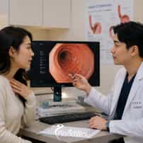
Upper GI Endoscopy in Gangnam, South Korea: Endoscopy for Reflux, Pain or Screening
Waking up with a burning sensation in the chest or a persistent metallic taste in the back of the throat is an exhausting way to start the day. For many individuals experiencing these symptoms, chronic upper digestive discomfort simply becomes a frustrating routine managed with daily over-the-counter antacids and cautious diet adjustments. Eventually, however, symptom management loses its effectiveness, and the need for a definitive, structural answer becomes impossible to ignore. The decision to undergo an upper gastrointestinal endoscopy, widely known as a gastroscopy, is rarely made on a whim. It usually follows months or even years of guessing what might be causing persistent abdominal pain, chronic acid reflux, or alarming difficulty swallowing. When long wait lists at home or prohibitively high out-of-pocket costs complicate access to this vital diagnostic tool, many patients begin researching international options, frequently discovering the comprehensive health screening centers in Gangnam, South Korea. The Daily Reality of Upper GI Symptoms The human upper digestive tract is a complex, highly sensitive system designed to move food smoothly while protecting the rest of the body from highly corrosive stomach acids. When this system begins to malfunction, the resulting symptoms can range from a mild, annoying ache to severe, sharp chest pain that mimics a cardiac event. Because the esophagus lacks a protective mucosal layer comparable to the stomach, prolonged exposure to acid causes significant inflammation, leading to a condition known as esophagitis. When to Stop Waiting: Recognizing Alarm Symptoms Professional gastroenterology guidelines generally recommend an upper GI endoscopy when symptoms do not respond to initial medical therapy, or when specific diagnostic red flags appear. These "alarm symptoms" may include unexplained weight loss, difficulty or pain when swallowing, evidence of gastrointestinal bleeding, persistent vomiting, or a direct family history of upper digestive cancers. If these signs are part of your situation, formal diagnostic evaluation should take priority over continued symptom masking. For those who have reached this threshold, the primary emotion is often a mixture of exhaustion and anxiety. The physical discomfort is draining, but the psychological burden of not knowing the exact cause is often worse. Patients frequently cycle through various primary care visits, receiving different empirical prescriptions without ever seeing the internal reality of their own anatomy. The Anatomical Pathway To understand why this procedure is so valuable, it helps to map the anatomy the scope actually travels through. The upper digestive tract is divided into three distinct zones, each vulnerable to its own specific conditions: The Esophagus: The muscular tube that relies on rhythmic contractions to propel food downward, highly susceptible to acid burns from a weakened lower sphincter. The Stomach: A highly acidic environment lined with a robust mucosal barrier that can sometimes be compromised by common bacteria or prolonged use of anti-inflammatory drugs. The Duodenum: The first part of the small intestine, acting as the primary mixing chamber for stomach contents and digestive enzymes, where peptic ulcers frequently form. The Limits of Empirical Treatment In many healthcare systems, the first line of defense for indigestion or reflux is empiric therapy. This involves prescribing acid-reducing medications, such as proton pump inhibitors (PPIs), to see if the symptoms improve without formally diagnosing the underlying cause. While this approach is often effective for simple, temporary heartburn, it becomes highly problematic when utilized as a long-term solution for chronic pain. Diagnostic Approach What It Offers What It Misses Empiric Therapy (Guessing) Provides temporary symptom relief and tests a hypothesis by using acid-reducing medication. Cannot confirm structural causes, identify precancerous cellular changes, or detect active bacterial infections. Barium Swallow (Imaging) Highlights the outline of the digestive tract on an X-ray to show strictures or large hernias. Cannot show mucosal color changes, cannot detect microscopic inflammation, and cannot take tissue biopsies. Upper GI Endoscopy (Seeing) Provides direct high-definition visualization of the tissue and allows for immediate biopsy and intervention. Focuses primarily on structure rather than motility function; functional nerve disorders may require additional specific testing. By merely turning off the acid pumps in the stomach, medication can successfully mask the pain while allowing the actual disease process to continue unchecked. If a patient is simply treating the symptom, they remain completely blind to the microscopic changes occurring in their cellular tissue. This is a critical distinction for patients to understand when deciding whether to pursue a formal endoscopic evaluation. The Endoscopic Technology An endoscope is far more than a simple tube. Modern gastroscopes are highly sophisticated instruments equipped with high-definition digital sensors, powerful light sources, and internal channels that allow the physician to wash the tissue, inflate the stomach with air for a clearer view, and pass tiny instruments down into the digestive tract. This real-time feedback allows the endoscopist to meticulously examine the mucosal architecture for the slightest irregularities. Beyond standard white-light cameras, many advanced screening centers now utilize digital enhancement technologies. By altering the specific wavelengths of light used during the examination, doctors can highlight different layers of the tissue to catch abnormalities sooner. High-Definition White Light: Provides a natural color view of the mucosa, allowing the doctor to spot obvious ulcers, areas of active bleeding, and larger polyps. Narrow Band Imaging (NBI): Uses specific blue and green light wavelengths that are heavily absorbed by hemoglobin, creating stark visual contrast that makes microscopic blood vessels and early abnormal cell clusters stand out vividly. Chromoendoscopy: Involves applying specialized safe dyes to the stomach lining during the procedure to highlight abnormal tissue architecture, though digital NBI has largely replaced this in many modern clinics. This level of technological precision changes the entire nature of the procedure, evolving from a simple visual check into a comprehensive micro-mapping of the upper digestive system. However, a high-definition screen is only valuable if the specialist knows exactly what subtle patterns to look for, which is why diagnostic volume becomes a critical factor in medical travel. Why Gangnam? The South Korean Screening Ecosystem Geography matters in healthcare, particularly when it comes to diagnostic repetition. Historically, East Asia, and South Korea in particular, has experienced some of the highest rates of stomach cancer in the world. This public health challenge was largely driven by dietary factors, high sodium intake, and the prevalent spread of the Helicobacter pylori bacteria across the population. Evidence and Public Health Impact In response to high gastric cancer rates, South Korea launched one of the most aggressive and comprehensive national cancer screening programs globally, making routine gastroscopies a standard part of adult healthcare. Published research has consistently demonstrated that this widespread implementation of endoscopic screening significantly reduces gastric cancer mortality in the population. For a medical traveler visiting a health screening center in Gangnam, this data represents deep, systemic expertise. The gastroenterologists in these high-volume centers perform and analyze these exams constantly, honing their ability to detect subtle mucosal abnormalities that might be easily overlooked elsewhere. This unique environment makes Seoul, and specifically well-resourced medical districts like Gangnam, incredibly suited for patients seeking diagnostic clarity. Rather than waiting months for a referral back home, a patient can often access a state-of-the-art screening center that integrates advanced imaging, immediate biopsy capabilities, and expert interpretation into a single, seamless appointment. Confronting the Procedure: The 15-Minute Reality Despite knowing the value of the procedure, many patients delay booking their appointment due to fear. The idea of having a camera passed down the throat triggers a very primal vulnerability. People worry about gagging, a feeling of suffocation, or waking up during the examination. Acknowledging these fears is a necessary part of the patient journey, but breaking down the actual timeline helps shift the perspective from dread to practical preparation. Phase 1: The Fasting Period Because the stomach empties its contents relatively quickly, patients are generally only required to fast from solid foods for about six to eight hours before the appointment. Clear liquids may be restricted a few hours prior to the procedure. This ensures that the stomach is entirely empty, providing the camera with a clear view and eliminating the risk of aspiration during sedation. Phase 2: The Sedation Choice In South Korean screening centers, conscious or deep intravenous sedation is widely favored and frequently offered as the standard of care for international patients. Through a small IV, medication is administered that places the patient into a deep, peaceful sleep. They continue to breathe entirely on their own, but they have no awareness of the scope and typically wake up with no memory of the examination itself. Phase 3: The Examination Once the patient is comfortably sedated, a small bite block is placed between the teeth. The doctor gently guides the endoscope down the esophagus, inflating the stomach slightly with air to smooth out the internal folds for a comprehensive view. The entire visual journey through the stomach and duodenum usually takes only 10 to 15 minutes before the scope is removed. Immediate Answers: Biopsies and H. pylori One of the most profound advantages of an endoscopy is that it is not merely a passive observation; it is an active, investigative tool. If the gastroenterologist sees an area of inflammation, an unusual growth, or a patch of tissue that looks slightly altered under Narrow Band Imaging, they can immediately pass tiny forceps through the scope to collect a microscopic biopsy. Because the lining of the digestive tract does not have pain receptors in the same way skin does, taking a biopsy causes no localized pain to the patient. Helicobacter Pylori Testing This common bacteria burrows into the stomach lining, causing chronic inflammation and vastly increasing the risk of ulcers and gastric cancer. A rapid urease test performed on a biopsy sample during the endoscopy can confirm its presence almost immediately, allowing doctors to prescribe a targeted antibiotic eradication protocol. Barrett’s Esophagus Monitoring For patients with years of chronic acid reflux, the cells in the lower esophagus can mutate to resemble intestinal cells in an attempt to protect themselves from the acid. A biopsy is the definitive way to confirm this precancerous condition, which requires careful, routine monitoring. Hearing that a biopsy was taken can cause a brief moment of panic for patients when they wake up. It is vital to understand that in the context of upper digestive health, biopsies are routine. They are frequently used to confirm benign conditions, check for microscopic healing, or test for common bacterial infections, rather than solely hunting for malignancies. The Financial Landscape of Screening in Seoul When evaluating medical travel for an endoscopy, price transparency helps frame the decision. In Gangnam, the cost for an upper GI endoscopy generally ranges from $300 to $2,000. This wide variance is not typically based on the quality of the camera, but rather on whether the procedure is included in a broader health screening package in Gangnam, South Korea. Country Typical Price (USD) Why Patients Travel From Here South Korea (Gangnam) $300 – $2,000 The baseline price referenced throughout this article. United States $1,500 – $4,000 Upper GI endoscopy in the US is commonly billed as a facility procedure, which pushes cash-pay pricing well above Korea's range even for routine screening. Russia $1,000 – $3,500 Internal-medicine screening is one of the categories where Russian patients most consistently travel to Korea, per Korean medical-tourism reporting. Mongolia $800 – $2,800 Mongolian patients are a recognized source market for Korean internal medicine and screening services, reflecting more limited advanced endoscopy access at home. Figures are general market ranges compiled from multiple clinics and medical-travel sources as of 2026; individual quotes vary by clinic, technique, and case complexity. Understanding what drives this pricing spectrum helps patients align their booking with their actual diagnostic needs: The Standalone Diagnostic ($300 - $600): Patients are typically paying for a standalone gastroscopy with conscious sedation. This usually includes the pre-consultation, the procedure, and a basic review of the visual findings. Biopsy pathology fees may occasionally be billed separately if tissue is actually sent to the lab. The Comprehensive Health Checkup ($1,000 - $2,000): Higher-tier prices almost always indicate that the endoscopy is bundled into a comprehensive, multi-day health screening package. These packages frequently include a full colonoscopy, comprehensive blood panels, abdominal ultrasounds, and cardiac stress tests. Translation and Concierge Services: Specialized international clinics embed the cost of a dedicated medical interpreter, translated medical records, and logistical coordination directly into their fees, ensuring a frictionless experience for travelers. For many patients coming from regions with highly privatized medicine, this spectrum represents significant value, especially when factoring in the speed of access. The ability to secure a comprehensive screening without fighting through months of tiered referrals is a major logistical advantage. Travel Logistics and Continuity of Care Undergoing a medical procedure abroad is different from a standard vacation, even for something as routine as a gastroscopy. The primary variable that changes the travel experience is the use of sedation. The medication used to ensure patient comfort takes time to fully metabolize, meaning the 24 hours surrounding the appointment require a dedicated focus on rest rather than tourism. Travel Planning for the 24-Hour Window Companion Responsibilities: The clinic will typically require a designated adult to help the patient navigate a taxi or transport back to their hotel. The companion should also be prepared to listen to post-procedure instructions, as the patient may be groggy immediately upon waking. Post-Procedure Diet: After waking up, the throat may feel slightly scratchy. Patients are usually advised to start with gentle, soft foods—like a warm Korean porridge (juk)—before returning to a normal diet. Heavy, spicy, or highly acidic foods should be avoided on the first day. Flight Timing: Medical teams universally advise against taking a long-haul flight immediately after receiving intravenous sedation. Patients should plan to rest in Seoul for at least 24 to 48 hours following the procedure to ensure the medication has cleared their system entirely. The final step in the medical travel journey is ensuring continuity of care. A diagnosis made in Seoul is only useful if it can be acted upon when the patient returns home. Reputable international screening centers in Gangnam will provide patients with high-quality digital copies of their endoscopic images, along with translated, formal medical reports detailing any biopsy findings so local doctors can prescribe appropriate care. The Decision to Stop Guessing Living with chronic upper digestive symptoms requires a tremendous amount of mental energy. The daily calculations regarding what to eat, when to sleep, and which medications to take eventually become exhausting. While empirical treatments and lifestyle changes are valid first steps, they cannot provide the structural certainty required to manage long-term health effectively. Choosing to undergo an upper GI endoscopy is a decision to prioritize clarity over comfort, and truth over assumption. For those who choose to pursue this clarity in Gangnam, the journey offers access to a healthcare ecosystem built on the principles of high-volume precision, advanced imaging technology, and streamlined preventative care. It transforms a daunting medical hurdle into a manageable, highly controlled step toward reclaiming a comfortable, ordinary life. Frequently Asked Questions How long does a gastroscopy procedure take? The actual endoscopic examination usually takes only 10 to 15 minutes. However, the entire appointment, including preparation, sedation administration, and recovery in the observation area, generally requires about two hours at the clinic. Will I be awake during the endoscopy? Most international patients in South Korea opt for conscious or deep sedation, meaning they are completely asleep or deeply relaxed during the procedure. Patients typically remember nothing about the examination itself and wake up comfortably in the recovery room. What is the difference between an endoscopy and a gastroscopy? Endoscopy is a broad medical term for any procedure that uses a lighted camera to look inside the body. A gastroscopy, also formally known as an upper GI endoscopy, is a specific type of endoscopy focused entirely on evaluating the esophagus, stomach, and duodenum. How soon can I fly after having an endoscopy in Seoul? If you receive sedation, clinical teams generally recommend waiting at least 24 hours before taking a long-haul flight to ensure the medication has fully cleared your system. If biopsies are taken, your doctor may suggest a slightly longer observation window depending on your specific situation. Does the procedure hurt? Because the procedure is typically performed under sedation, patients do not experience pain during the examination. Following the procedure, you may feel mild bloating from the air used to inflate the stomach, or experience a minor, temporary sore throat. Why are biopsies taken during the examination? Biopsies are routine samples of tissue used to check for microscopic inflammation, precancerous cellular changes (like Barrett's esophagus), or the presence of the Helicobacter pylori bacteria. Taking a biopsy is a standard diagnostic protocol and does not automatically mean cancer is suspected. Ready to Stop Guessing? Connect with comprehensive health screening centers in Gangnam to clarify your diagnosis, compare package options, and coordinate your medical travel schedule safely. Request Information References https://pubmed.ncbi.nlm.nih.gov/28109822/ https://pubmed.ncbi.nlm.nih.gov/36183307/ https://pubmed.ncbi.nlm.nih.gov/22624976/ https://pubmed.ncbi.nlm.nih.gov/26601217/ https://pubmed.ncbi.nlm.nih.gov/34353846/ https://pubmed.ncbi.nlm.nih.gov/34293997/ This content is for educational purposes only and is not medical advice. Diagnosis, treatment suitability, risks, recovery, travel timing and outcomes vary by individual and procedure. Decisions should be made with appropriately qualified healthcare professionals after an individual assessment. PlacidWay provides healthcare information and facilitation services and is not the treating physician, clinic or hospital....
Read more details

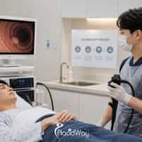
Colonoscopy in Gangnam, South Korea: Bowel Prep, Timing and Polyp Detection
For many adults, the decision to undergo a colonoscopy is marked by procrastination, preparation concerns, and anxiety about the results. When traveling to South Korea for screening, colonoscopy in Gangnam is often integrated into comprehensive health screening programs, making the process more streamlined and efficient. When individuals travel to South Korea for medical care, they often discover an entirely different approach to preventative medicine. In Seoul, and particularly in districts like Gangnam, a colonoscopy is rarely treated as an isolated, anxiety-inducing appointment. Instead, it is frequently integrated into highly efficient, comprehensive health screening programs, transforming a dreaded task into a streamlined component of a personal health audit. A Shift in Perspective Traveling for a colonoscopy changes the context of the procedure entirely. Rather than squeezing an appointment between work meetings, patients dedicating a week to health screening in Seoul can focus entirely on the preparation, the procedure, and their recovery. However, this shift in geography introduces new practical realities, such as managing fasting protocols in a foreign city and calculating exactly when it is medically safe to board a long-haul flight home. The Efficiency of the Korean Health Checkup Culture South Korea has developed one of the most robust preventative healthcare infrastructures in the world, built on a model of high-volume precision. Comprehensive health screening centers operate with remarkable synchronization, often dedicating entire floors of major hospitals or private Gangnam clinics to coordinated diagnostic testing. Patients move seamlessly from blood work to imaging, and finally to the endoscopy suite, all within a single morning schedule. This efficiency is not merely about saving time; it fundamentally changes the psychological experience of preventative care. By consolidating diagnostics into a single visit, clinics dramatically reduce the fragmented anxiety that usually accompanies medical testing. For international visitors, this means that a trip designed for a medical checkup can yield a complete physiological baseline in a matter of hours, rather than requiring weeks of separate appointments across different specialist offices. Consolidated Care Endoscopies are routinely paired with full cardiovascular, organ, and metabolic assessments during the same fasting period. This allows the medical team to maximize the utility of the patient's pre-procedure diet restrictions. Advanced Facilities Screening centers are designed specifically for asymptomatic individuals, prioritizing comfort, privacy, and rapid transit between testing stations. Patients are guided by dedicated concierges or digital wristbands to eliminate waiting room confusion. Same-Day Action If precancerous polyps are identified during the screening, they are typically removed immediately during the initial scope insertion. This proactive intervention prevents the need for a secondary procedure and provides immediate peace of mind. Western Fragmentation vs. Korean Integration Many medical tourists are primarily motivated by the structural bottlenecks they consistently face in their domestic healthcare systems. In numerous Western countries, scheduling a routine colonoscopy involves securing a referral from a primary care physician, attending a separate consultation with a gastroenterologist, and facing a subsequent wait for the actual procedure date. If a polyp is discovered and removed, pathology results may take several additional weeks, unnecessarily extending the period of clinical uncertainty. The Gangnam screening model contrasts this by functioning as a centralized, patient-directed diagnostic hub. Because these specialized facilities cater specifically to proactive preventative care, the traditional referral hurdles are entirely removed. Patients can book directly with the center, undergo the procedure alongside other diagnostics, and often sit down for a comprehensive review of their initial results with an English-speaking specialist on the exact same day. Experience Factor Traditional Fragmented Model Integrated Gangnam Model Access & Scheduling Often requires primary care referrals and waiting lists spanning weeks or months. Direct booking available for comprehensive packages, often scheduled within days. Diagnostic Scope Colonoscopy is performed as an isolated, standalone appointment. Frequently bundled with gastroscopy, full-body imaging, and advanced blood panels. Results Delivery Pathology and imaging reviews often require a follow-up appointment weeks later. Initial visual results are discussed same-day; pathology is typically expedited and digitally translated. The Hotel Reality: Managing Bowel Prep Abroad Regardless of where in the world a colonoscopy is performed, the clinical success of the procedure relies entirely on the quality of the patient's bowel preparation. If the colon is not completely clear of residue, the endoscopist cannot clearly see the mucosal lining, significantly increasing the risk of missing flat or small polyps. For international patients, managing this strict preparatory protocol while staying in a Gangnam hotel requires careful logistical planning and personal discipline. Modern gastroenterology guidelines strongly support a "split-dose" preparation regimen for optimal visualization. Research consistently shows that taking half of the purgative liquid the evening before the procedure, and the remaining half early on the morning of the procedure, results in a significantly cleaner colon and is generally easier for patients to tolerate. Executing this specific timeline in a hotel room means setting early alarms and ensuring immediate, uninterrupted access to a private en-suite bathroom. ! The Diet Challenge in Seoul Seoul is internationally famous for its vibrant, highly seasoned, and fiber-rich cuisine. However, for three days prior to a colonoscopy, patients must strictly adhere to a low-residue diet, making it necessary to actively avoid the very foods the city is known for. What to strictly avoid: Kimchi, sesame seeds, multi-grain rice, seaweed (gim), fresh vegetables, and spicy red pepper paste (gochujang). What is generally safe: White rice (bap), plain white bread, clear broths, soft tofu, eggs, and steamed white fish. Clear liquid day (24 hours prior): Local convenience stores are excellent resources for approved clear fluids like Pocari Sweat (a clear Korean sports drink), apple juice, and plain bottled water. When checking into a hotel before the procedure, it is highly advisable to familiarize yourself with the room's specific amenities. Ensuring you have adequate refrigeration for the prep liquid—which is often much more palatable when consumed chilled—and knowing the location of the nearest 24-hour convenience store can relieve significant pre-procedure stress. The chosen clinic will usually provide the physical preparation kit during an initial pre-consultation, or arrange for it to be securely delivered directly to your accommodation. International coordinators at major Gangnam clinics are accustomed to explaining these steps in English, but the crucial responsibility of consuming the exact required volume of liquid ultimately falls to the patient alone in their hotel room. The Science of Seeing: Why Equipment and Detection Rates Matter Patients frequently assume that a colonoscopy is a universally standardized commodity, believing that having the procedure done anywhere yields the exact same diagnostic accuracy. This is a critical misunderstanding of gastroenterological science. The ability to locate subtle, precancerous growths depends heavily on the specific generation of technology used and the highly specialized observational skill of the physician operating the scope. Top-tier clinics in South Korea for colonoscopy frequently utilize high-definition, narrow-band imaging (NBI) scopes that optically highlight abnormal vascular patterns in the mucosal lining. Several advanced facilities have also integrated artificial intelligence (AI) detection systems, which act as a digital second set of eyes by actively flagging subtle color changes or flat lesions on the monitor. However, even the most sophisticated technology alone does not guarantee a high-quality screening outcome. Understanding the Adenoma Detection Rate (ADR) The absolute most important quality indicator for a screening colonoscopy is the Adenoma Detection Rate (ADR). This metric represents the exact percentage of screening colonoscopies in which the physician successfully finds one or more precancerous polyps (adenomas). Current medical guidelines from major gastrointestinal societies suggest an ADR target of at least 25% to 35% for endoscopists, depending on patient demographics. Clinical research strongly indicates that for every 1% increase in a physician's ADR, the patient's individual risk of developing interval colorectal cancer drops by approximately 3%. When evaluating clinics in Seoul, the conversation should naturally move beyond simple hotel packages and focus heavily on whether the facility tracks and meets internationally recognized ADR benchmarks. Achieving a consistently high ADR requires a meticulous and disciplined procedural approach from the physician. The endoscopist must take adequate time during the crucial withdrawal phase of the scope—typically dedicating a minimum of 6 to 9 minutes—to carefully inspect behind every anatomical fold of the colon. A rushed procedure, regardless of how advanced the camera system might be, dramatically lowers the clinical effectiveness of the screening. For medical tourists evaluating their options, this means that selecting a facility should never be based solely on package pricing or aesthetic clinic appeal. The true clinical value of traveling to Gangnam for a colonoscopy lies in deliberately accessing high-volume specialists who operate with top-tier optical equipment and dedicate the necessary time to thorough mucosal inspection. Anatomy of Polyps: What Doctors Look For Understanding what physicians are actually looking for helps demystify the importance of advanced, high-definition imaging. Polyps are abnormal growths of tissue projecting from the mucous membrane of the colon, and while the majority of these growths are benign, a specific subset carries the genetic potential to slowly transform into colorectal cancer over several years. Identifying the precise structural type of a polyp during the procedure directly dictates how aggressively the endoscopist will act. Because distinguishing between a harmless growth and a high-risk lesion visually requires immense precision, the clarity provided by Gangnam's high-definition diagnostic monitors becomes a crucial clinical asset. Adenomatous Polyps These are the most common type of precancerous polyps found during routine screenings. They carry a distinct risk of turning into cancer if left to grow unchecked over a period of several years. Hyperplastic Polyps These are typically harmless growths that present a near-zero risk of developing into malignancy. However, they must still be carefully examined by pathology to confirm they are entirely benign. Sessile Serrated These are flat, subtly colored lesions that blend seamlessly into the colon wall. This makes them notoriously difficult to detect without high-definition optical enhancement or specialized dye sprays. Inside the Procedure Room: The "Sleep Endoscopy" Experience A defining feature of the Korean health screening experience is the widespread, standard use of what local clinics refer to as "sleep endoscopy" (sumyeon naesigyeong). While medical practices in some Western countries still rely heavily on conscious sedation—where the patient remains groggy but partially awake and responsive—Korean facilities heavily favor deep sedation protocols for gastrointestinal screenings. Under sleep endoscopy, patients are rendered completely unaware of the clinical environment and the procedure itself. Medications such as propofol or midazolam are administered intravenously by the medical team, inducing a rapid, highly controlled deep sleep. The patient typically wakes up in a dedicated recovery bay 30 to 45 minutes later with absolutely no memory of the scope insertion, inflation, or any polyp removal. Safety and Monitoring Protocols Because deep sedation inherently carries minor respiratory risks, reputable screening centers in Gangnam employ dedicated anesthesiologists or highly specialized monitoring staff. Continuous pulse oximetry, automated blood pressure tracking, and oxygen partial pressure measurements are standard during the procedure to ensure the patient's airway and vitals remain entirely stable while the endoscopist focuses entirely on the screening. Waking up from sleep endoscopy is usually a swift process, but a mandatory monitored recovery period is strictly enforced by the clinic. Patients are escorted to a quiet, dimly lit resting area where vital signs are tracked until the medication fully wears off. Because residual grogginess can subtly impair judgment and reflexes, clinics strictly prohibit patients from driving or operating heavy machinery for the remainder of the calendar day. For international travelers navigating a foreign city, this strictly enforced recovery protocol underscores the immense practical importance of having a travel companion. While it is physically possible to take a Seoul taxi back to a hotel alone, having someone to assist with translation, physically handle discharge instructions, and ensure a safe, smooth return to your accommodation provides critical peace of mind. The "What If": Polyp Detection and Removal Abroad The primary clinical goal of a screening colonoscopy is not just passive diagnosis, but active cancer prevention. If a physician spots an abnormal polyp forming on the mucosal lining of the colon, they will almost always elect to remove it immediately during the very same procedure. This proactive intervention is exactly what makes a colonoscopy uniquely powerful; it physically stops potential cancer before it has the opportunity to form and spread. However, having a polyp surgically removed while overseas introduces an important layer of logistical complexity to a travel itinerary. A polypectomy is fundamentally a minor surgical procedure performed inside the bowel wall. While the colon lacks internal pain receptors to physically feel the cutting or thermal burning of the tissue, a small internal wound is left behind to slowly heal over the following weeks. This internal healing process strictly dictates what patients can safely do in the immediate days following their appointment. Pathology and Result Translations Once the tissue is successfully removed, it must be carefully sent to a specialized pathology laboratory to determine if it was benign, a precancerous adenoma, or frankly malignant. In highly efficient Korean health centers, these microscopic results can sometimes be expedited for international patients, but they generally require a few dedicated business days to properly process and report. Patients must proactively plan for exactly how they will receive and comprehend these specialized medical results. If your scheduled flight departs before the official pathology report is finalized, you need absolute certainty that the clinic provides secure, medically translated digital results via email or a patient portal. Understanding whether a removed polyp was an adenoma or a harmless hyperplastic growth dictates when your next colonoscopy should be scheduled—a vital piece of clinical information you must hand over to your home physician. The Altitude Factor: Flying Safely After a Polypectomy The most critical travel restriction directly associated with a colonoscopy involves commercial air travel. While patients can usually return to their normal ground-level activities and diet the following day, stepping onto a pressurized passenger aircraft requires very careful medical consideration. This is particularly true if any form of surgical intervention or tissue removal occurred during the screening. The physiological danger of flying after bowel surgery lies in fundamental physics and gas expansion. At commercial cruising altitude, the cabin pressure noticeably drops, causing any residual gases trapped naturally inside the gastrointestinal tract to physically expand in volume. This expansion directly stretches the delicate healing mucosal lining where a polyp was recently removed. In severe cases, this pressure-induced stretching can induce delayed hemorrhage (bleeding) or, very rarely, a life-threatening perforation of the bowel wall. Procedure Outcome General Flight Guideline Medical Reasoning Routine Scope (No Polyps) Usually safe after 24 hours Allows deep sedation to fully clear and residual endoscopic gases to naturally pass. Small Polyp Removal Requires clearance; often 48 to 72 hours minimum Allows the initial mucosal wound to stabilize against the risk of pressure-induced stretching. Large or Complex Polyp Removal May require 7 to 14 days of travel delay Delayed bleeding risk peaks between days 5 and 7. Experiencing a GI bleed mid-flight is a severe emergency. Because the presence, quantity, and exact size of polyps are entirely unpredictable before the endoscope is actually inserted, medical tourists must intentionally build buffer days into their Seoul itinerary. Scheduling a colonoscopy for the afternoon before a major trans-Pacific flight is a highly dangerous logistical gamble. If a surprisingly large polyp is discovered and removed, the treating physician may medically forbid you from flying, forcing last-minute flight changes, penalty fees, and extended hotel stays. To effectively mitigate this financial and medical risk, patients are strongly advised to schedule their health screenings at the very beginning of their trip to South Korea. This strategic scheduling provides a natural, stress-free recovery window while enjoying the cultural sights of the country. It ensures that if an intervention occurs, adequate time safely passes before exposing the delicate healing bowel to severe altitude and pressure changes. Understanding the Financials: The $300 to $2,000 Range The overall cost of healthcare in South Korea is highly competitive on a global scale, but the wide price range for colonoscopies—from roughly $300 to $2,000 USD—can cause significant confusion for researching international patients. This financial variance is rarely about the baseline quality of the endoscope or the physician's credentials. Instead, it directly reflects how the specific procedure is packaged and what exact supplementary diagnostic services are included in the final bill. Country Typical Price (USD) Why Patients Travel From Here South Korea (Gangnam) $300 – $2,000 The baseline price referenced throughout this article. United States $1,500 – $4,000 A cash-pay colonoscopy in the US typically costs several times Korea's range once facility and anesthesia fees are included. Russia $1,000 – $3,500 Colon cancer screening is a frequently cited reason Russian patients travel to Korea for internal medicine care. Mongolia $800 – $2,800 Screening colonoscopy is among the procedures driving Mongolian patient travel to Korea's internal medicine departments. Figures are general market ranges compiled from multiple clinics and medical-travel sources as of 2026; individual quotes vary by clinic, technique, and case complexity. At the lower end of this financial spectrum (closer to $300), patients are typically paying for a standalone screening colonoscopy under standard sedation at a highly specialized local gastroenterology clinic. This base price generally covers the initial consultation, the prescribed prep kit, the procedure itself, and the standard sedation medications. However, if multiple complex polyps are unexpectedly found, specialized pathology lab fees and surgical removal fees may be billed as additional on-site costs at checkout. Comprehensive Screening Packages The much higher end of the price spectrum represents comprehensive, executive-level health checkup packages at elite Gangnam diagnostic centers or major university hospital wings. When a patient pays $1,500 to $2,000, the colonoscopy is just one single piece of a much larger, highly coordinated diagnostic puzzle. These premium packages routinely bundle the colonoscopy with an upper endoscopy (gastroscopy), full-body MRI or low-dose CT scans, extensive cardiovascular stress testing, comprehensive tumor marker blood panels, and dedicated English-speaking concierge services from arrival to departure. When evaluating these varied costs, international patients must explicitly clarify exactly what is legally included in their quoted price. It is critical to confirm whether deep sedation fees are built into the base price, how sudden polyp removal is structurally billed, and if formal English translation of the final medical report is provided entirely free of charge or requires an extra administrative fee. Continuity of Care: What Happens When You Return Home A truly successful international health screening does not end the moment you walk out of the Seoul clinic; it formally ends when your primary care physician back home comprehensively understands the results. The highly detailed physiological data gathered during a Gangnam checkup is only clinically useful if it can be seamlessly integrated into your long-term, permanent health record. Before departing South Korea, you must ensure you have fully secured high-resolution digital copies of all internal imaging and official, certified English translations of any pathology reports. If polyps were surgically removed, the exact size in millimeters, precise location within the colon, and complete histology (the cellular makeup) must be documented in writing. This specific information is critical because it directly determines the strict medical guidelines for your next screening—whether you are safely cleared for ten years, or need a rigorous follow-up in just three years. Questions for Your Gangnam Clinic Coordinator Will I receive the bowel prep kit at my hotel, and are the precise dosing instructions provided clearly in English? What is the clinic’s specific policy and billing structure if a polyp unexpectedly needs to be removed during the procedure? How many days does the clinic require me to legally stay in Seoul before authorizing air travel if a polypectomy occurs? Are the final pathology results formally translated into English, and how are they delivered if I have already flown home? Does the facility actively monitor the Adenoma Detection Rate (ADR) of its endoscopists, and are those rates publicly shared? Undergoing a comprehensive colonoscopy in a foreign, bustling city introduces undeniable logistical layers that patients simply do not face at home. The reality of properly fasting in a commercial hotel, meticulously timing a long-haul flight, and successfully navigating an entirely different medical culture requires serious preparation and foresight. Yet, for many proactive travelers, the extreme efficiency, top-tier technology, and highly comprehensive nature of a Gangnam health screening provide the exact motivation needed to finally stop procrastinating. By deeply understanding the specific travel risks, carefully preparing for the rigid dietary realities, and knowing exactly what questions to ask coordinators in advance, patients can genuinely reclaim control of their health timeline. This proactive approach ultimately replaces the anxiety of the unknown with clear, actionable medical insights. Frequently Asked Questions How do I complete the bowel prep in a hotel room? Bowel prep involves drinking a split-dose purgative liquid. You will need access to a private bathroom and refrigeration for the fluids. You must avoid fiber-rich and spicy foods for a few days prior, relying on clear liquids available at local convenience stores on the day before the procedure. What is "sleep endoscopy" in South Korea? Sleep endoscopy uses deep sedation (often propofol or midazolam) rather than conscious sedation. Patients are completely asleep during the procedure and wake up in a recovery bay with no memory of the screening, monitored closely by medical staff. How soon can I fly home after a colonoscopy in Seoul? If no polyps are found, flying after 24 hours is generally safe. If polyps are removed, you must delay flying due to the risk of cabin pressure causing delayed bleeding. Small polyps may require 48-72 hours, while larger polyp removal might demand a 7 to 14-day delay before air travel. Are polyps removed immediately if found? Yes, in most cases, if a physician identifies a polyp during the colonoscopy, they will perform a polypectomy to remove it immediately. The tissue is then sent for pathological analysis to determine your risk level. Why is there a wide price range for colonoscopies in Gangnam? Prices range from roughly $300 for a standalone colonoscopy at a specialized clinic to $2,000 for a comprehensive health screening package. These premium packages often include cardiovascular testing, MRI scans, upper endoscopies, and dedicated concierge services. What is the Adenoma Detection Rate (ADR) and why does it matter? The ADR is the percentage of colonoscopies where a doctor successfully finds precancerous polyps. Current medical guidelines suggest an ADR target of at least 25% to 35%, as a higher detection rate is directly linked to a significantly lower risk of interval colorectal cancer. Plan Your Health Screening with Clarity If you need assistance comparing health screening packages, understanding protocol details, or communicating with specialists in Seoul, our team can help you navigate the next steps. We can provide objective guidance based on your specific medical requirements. Request Information References https://pubmed.ncbi.nlm.nih.gov/36638814/ https://pubmed.ncbi.nlm.nih.gov/24011871/ https://pubmed.ncbi.nlm.nih.gov/38383266/ https://www.ncbi.nlm.nih.gov/pmc/articles/PMC7341512/ https://www.ncbi.nlm.nih.gov/pmc/articles/PMC3991959/ https://www.ncbi.nlm.nih.gov/pmc/articles/PMC6453579/ https://www.ncbi.nlm.nih.gov/pmc/articles/PMC8248943/ https://pubmed.ncbi.nlm.nih.gov/25597420/ This content is for educational purposes only and is not medical advice. Diagnosis, treatment suitability, risks, recovery, travel timing and outcomes vary by individual and procedure. Decisions should be made with appropriately qualified healthcare professionals after an individual assessment. PlacidWay provides healthcare information and facilitation services and is not the treating physician, clinic or hospital....
Read more details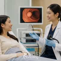
Endoscopic Polyp Removal in Gangnam, South Korea: Same-Session Polyp Treatment
You schedule an endoscopy for peace of mind, but discovering a polyp can quickly raise concerns about cancer risk and further procedures. With endoscopic polyp removal in Gangnam, selected polyps may be removed during the same session, potentially avoiding a second procedure and another round of bowel preparation. The Core Decision: Diagnostic vs. Therapeutic Screening The profound relief of waking up to hear, "We found a polyp, and we have already removed it," is the primary reason international patients seek out specialized screening centers. Same-session therapeutic treatment eliminates the agonizing wait between discovery and intervention, fundamentally changing the psychological and practical experience of preventive care. Understanding Polyps and Endoscopic Removal A polyp is an abnormal growth of tissue projecting from the mucosal lining of the gastrointestinal tract, most commonly found in the colon or the stomach. While the word itself often causes immediate fear, the vast majority of these growths are entirely benign and take years to develop into anything medically dangerous. Endoscopic removal, clinically known as a polypectomy, is the definitive preventive measure that severs and retrieves the abnormal tissue before it can undergo malignant changes. During the procedure, a flexible tube equipped with a high-definition camera and precise lighting is navigated slowly through the digestive tract. When an anomaly is spotted on the monitor, the gastroenterologist passes specialized micro-instruments through a hollow channel in the endoscope to safely detach the tissue from the healthy organ wall. The severed tissue is then carefully withdrawn and sent for precise laboratory analysis to determine its exact cellular composition and confirm that the margins are clear. Hyperplastic Polyps These are very common, typically small growths that carry almost no risk of turning into cancer. They are often removed simply to confirm their benign nature under a microscope. Adenomatous Polyps Considered precancerous, adenomas have the potential to become malignant over several years if left untouched. Identifying and removing them is the primary goal of routine colonoscopies. Malignant Polyps These are growths that already contain cancerous cells. Depending on the depth of cellular invasion, endoscopic removal might be sufficient, or subsequent surgery may be required. The Reality of Bowel Preparation and Fasting Regardless of where in the world an endoscopy is performed, the safety and accuracy of the procedure rely entirely on the strict quality of the patient's preparation. For a colonoscopy, this means adhering strictly to a clear liquid diet and consuming a specialized bowel-cleansing solution the evening before and the morning of the exam. For a gastroscopy, it requires a complete fasting period to ensure the stomach is entirely empty, allowing the high-definition cameras an unobstructed view of the mucosal lining. Traveling internationally adds a unique layer of complexity to this preparation phase, as patients must navigate unfamiliar pharmacies, hotel accommodations, and local dietary options. Many premium screening centers in South Korea alleviate this burden by couriering the exact preparation medications directly to the patient's hotel, complete with detailed multilingual instructions. Having a dedicated resting space with a private bathroom is absolutely non-negotiable during the final twenty-four hours leading up to the appointment. Techniques: From Standard Snare to Mucosal Dissection Not all gastrointestinal lesions are identical, and the method used to extract them depends heavily on their precise size, shape, and depth within the tissue layers. Small, raised polyps that resemble tiny mushrooms are generally straightforward to capture and remove, while flat or broad-based lesions require a significantly higher level of technical expertise to lift away safely. The specific technique chosen by the gastroenterologist dictates both the overall safety of the procedure and the likelihood of the polyp returning in the future. When growths are larger than a few millimeters or spread flat against the intestinal lining, standard wire loops are often insufficient, and therapeutic techniques become strictly necessary. This is where the distinction between different screening facilities becomes medically critical, as not all global centers are equipped or staffed to perform complex mucosal resections during a routine morning checkup. Understanding the difference between these interventions helps clarify why certain clinics require highly specialized physicians. Removal Technique How It Works Best Suited For Complexity Level Standard Snare Polypectomy A thin wire loop is placed around the base of the polyp and tightened to cut the tissue, sometimes using mild electrocautery to seal vessels. Small, raised, or pedunculated (stalked) polyps easily visible during a routine scan. Low. Standard practice in nearly all global screening centers. Endoscopic Mucosal Resection (EMR) Fluid is injected under the polyp to lift it away from the deeper muscle wall, creating a safe cushion before a snare removes the raised tissue. Larger polyps (typically over 10-15mm) or flat lesions confined to the superficial mucosal layer. Moderate to High. Requires specialized training to ensure the entire lesion is removed cleanly. Endoscopic Submucosal Dissection (ESD) After fluid injection, a specialized microscopic knife is used to painstakingly carve the lesion away from the submucosal layer in one single piece. Complex, very large, or heavily flat lesions that carry a high suspicion of early-stage, localized cancer. Very High. A highly specialized skill requiring dedicated therapeutic endoscopy facilities. Why Gangnam? The Culture of Comprehensive Diagnostics When international patients search for healthcare options in Gangnam, they are often anticipating the district's famous aesthetic clinics, but the area is equally renowned for its hyper-efficient, technology-equipped health screening centers. Facilities such as those affiliated with major university hospitals operate massive diagnostic hubs designed specifically to evaluate the entire body in a single morning. This distinct environment has fostered a standard of care where finding and treating gastrointestinal issues happens seamlessly within the same visit, without administrative delays. The medical philosophy in these centers prioritizes immediate therapeutic intervention over prolonged diagnostic staging. Because gastric and colorectal cancers have historically been significant public health concerns in South Korea, the national medical infrastructure has heavily invested in early detection programs and endoscopic training. As a result, the gastroenterologists performing these screenings are typically high-volume specialists who encounter and successfully remove complex lesions daily. The Same-Session Workflow High-Definition Visualization: Centers utilize high-definition scopes and Narrow Band Imaging (NBI), which uses specialized light wavelengths to highlight the microscopic vascular structure of the tissue, making flat anomalies much easier to detect. Immediate Assessment: Once a polyp is identified, the endoscopist instantly evaluates its size, border, and surface pattern to determine if it can be safely removed on the spot. On-the-Spot Intervention: Rather than withdrawing the scope, the physician switches to therapeutic instruments, performing a snare polypectomy, EMR, or occasionally an ESD while the patient remains comfortably sedated. Post-Procedure Clarity: The patient wakes up in recovery and is informed of the removal, eliminating the need to schedule a separate surgical date and immediately beginning the pathology waiting period. Navigating the Evidence: Risks and the Travel Reality While endoscopic removal is overwhelmingly safe and minimally invasive, it is still a surgical procedure performed inside the body, and it carries distinct statistical risks that must be carefully planned for. The two primary clinical concerns following any polypectomy are delayed bleeding at the internal resection site and, far more rarely, microscopic perforation of the gastrointestinal wall. The likelihood of these events increases substantially if the removed polyp was exceptionally large, deeply rooted, or located in the thinner right side of the colon. According to published clinical literature, the overall risk of post-polypectomy bleeding across all procedures is generally around one percent, but this figure can rise toward five to ten percent for complex lesions removed via endoscopic mucosal resection. Perforation is highly uncommon, typically occurring in less than one in a thousand standard cases, though the risk profile changes appropriately with submucosal dissections. Understanding these variables is not meant to cause alarm, but rather to inform how long a patient must realistically remain near their treating facility before boarding a long-haul flight home. Crucial Travel Implication: Flying After Polypectomy For healthcare travelers, the greatest risk of delayed bleeding occurs between days three and seven after the procedure. The atmospheric pressure changes inside an airplane cabin can theoretically disrupt the delicate scab forming over the internal resection site. If a routine screening finds nothing, patients can generally fly within 24 hours. However, if a large polyp is removed using EMR or ESD techniques, medical guidelines often strongly suggest delaying commercial air travel for one to two weeks. You must build flexibility into your Seoul travel itinerary in case an unexpected intervention requires a longer recovery stay. The Caregiver’s Role and Continuity of Care Traveling for a medical screening frequently involves a companion, and their supportive role becomes unexpectedly vital if a routine diagnostic checkup turns into a therapeutic procedure. Because the patient will be slowly recovering from systemic sedation, they will not be legally or physically permitted to navigate Seoul’s busy transportation system or make complex medical decisions immediately after discharge. The companion must be fully prepared to receive the postoperative instructions, monitor the patient’s initial dietary intake, and understand the warning signs of potential complications. Once the initial recovery period has safely passed, the focus shifts to long-term continuity of care and managing the official pathology results. Even if a removed polyp is completely benign, its specific cellular type will dictate exactly when the patient needs their next colonoscopy or gastroscopy, which could be anywhere from one to ten years later. Ensuring that the South Korean medical team provides all imaging data, operative reports, and accurately translated pathology findings is crucial so that the patient's local doctor back home can seamlessly take over their future surveillance. Companion Checklist for Discharge Transportation: Securing a taxi or private transfer back to the hotel, as the patient cannot walk long distances or take complex public transit post-sedation. Dietary Management: Ensuring access to soft, easily digestible foods (such as Korean rice porridge, known as juk) for the first 24 to 48 hours to protect the healing mucosa. Emergency Contacts: Keeping the direct hotline for the screening center’s international desk readily available in the event of unexpected abdominal pain or bleeding. Record Collection: Verifying that all digital imaging (often provided on a USB or secure portal) and English-translated pathology reports are secured before leaving the country. A Clearer Path Forward Deciding to undergo a comprehensive health screening abroad is fundamentally about taking proactive control over your personal baseline of health. The true value of choosing a destination specifically equipped for same-session treatment lies in the profound peace of mind it offers, ensuring that if an anatomical anomaly is found, it is addressed immediately and skillfully. By eliminating the agonizing wait times and fragmented care models common in other systems, patients can return home with definitive answers rather than lingering, anxiety-inducing questions. Ultimately, the overarching goal of any endoscopic procedure is to intercept disease well before it has the opportunity to develop into a systemic threat. Whether you choose to incorporate a screening into a broader leisure trip to Seoul or travel specifically to access the specialized gastroenterology infrastructure of Gangnam, the most critical step is simply ensuring the evaluation takes place. With careful logistical planning, clear communication with the treating facility, and an understanding of the necessary recovery windows, you can confidently navigate this vital aspect of preventive medicine. Frequently Asked Questions What is the difference between a screening endoscopy and a therapeutic endoscopy? A screening endoscopy is primarily diagnostic, meant to visually inspect the gastrointestinal tract and perhaps take small biopsies. A therapeutic endoscopy involves active treatment during the same session, such as removing a discovered polyp using a snare, EMR, or ESD technique while the patient remains sedated. How long do I need to stay in South Korea after a polyp is removed? The required stay depends heavily on the size of the polyp and the removal technique used. While a standard screening with no findings allows for normal travel almost immediately, the removal of a large polyp via EMR or ESD may require you to delay flying for one to two weeks due to the risk of delayed bleeding associated with cabin pressure changes. Are all polyps cancerous? No, the vast majority of polyps are benign. However, certain types, such as adenomatous polyps, are considered precancerous and have the potential to develop into cancer over several years if they are not removed. Endoscopic removal is the primary preventive measure to ensure they do not become malignant. Does polyp removal hurt? Because the mucosal lining of the gastrointestinal tract lacks pain receptors for cutting or burning, the actual removal of a polyp is generally painless. Patients are typically under conscious sedation or deep sleep during the procedure and may only experience mild bloating or cramping afterward due to the air introduced into the tract for visibility. Why do patients travel to Gangnam for routine health screenings? Gangnam is home to numerous high-volume health screening centers that specialize in hyper-efficient, comprehensive evaluations. Patients travel there for the ability to undergo whole-body diagnostics, including highly specialized same-session endoscopic treatments, utilizing high-definition imaging technologies in a streamlined timeframe. Ready to Explore Your Screening Options? Compare comprehensive health screening packages, clarify clinical protocols, and connect with specialized endoscopic centers to plan your preventive care journey. Request Information References https://pubmed.ncbi.nlm.nih.gov/34450667/ https://pubmed.ncbi.nlm.nih.gov/22089297/ https://www.ncbi.nlm.nih.gov/pmc/articles/PMC8706313/ https://pubmed.ncbi.nlm.nih.gov/24477363/ https://www.ncbi.nlm.nih.gov/pmc/articles/PMC11710915/ https://pubmed.ncbi.nlm.nih.gov/25839689/ This content is for educational purposes only and is not medical advice. Diagnosis, treatment suitability, risks, recovery, travel timing and outcomes vary by individual and procedure. Decisions should be made with appropriately qualified healthcare professionals after an individual assessment. PlacidWay provides healthcare information and facilitation services and is not the treating physician, clinic or hospital....
Read more detailsDiscover your treatment options with a free, no-obligation quote!
Get your quote now!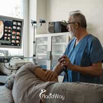
ESD Endoscopic Resection in Gangnam, South Korea: ESD for Early GI Lesions
The journey toward Endoscopic Submucosal Dissection (ESD) rarely begins with the intention to seek complex treatment. It typically starts with a routine screening, a follow-up endoscopy for persistent indigestion, or a colonoscopy designed to prevent problems rather than find them. When the physician calls to discuss the biopsy results, the initial diagnosis of an early gastrointestinal lesion or high-grade dysplasia often brings a confusing mix of immense relief and sudden clinical tension. When screening reveals an early gastrointestinal lesion or high-grade dysplasia, patients may face difficult treatment decisions. For selected cases, ESD endoscopic resection in Gangnam offers a minimally invasive option for removing early GI lesions. The Decision Fork: Efficacy Versus Organ Preservation Patients often find themselves caught between two highly undesirable extremes. They can opt for standard Endoscopic Mucosal Resection (EMR), which might require taking the lesion out in multiple fragments and risking recurrence, or they can choose open or laparoscopic surgery, which guarantees the removal of the disease but permanently alters their digestive anatomy. ESD represents a highly technical middle path—allowing the entire lesion to be peeled away from the inside without removing the organ itself. Recognizing that ESD exists is only the first step in the patient's research process. The subsequent realization is that this procedure demands a high level of technical skill, a steep learning curve, and an environment characterized by high procedural volume. This realization frequently prompts patients to look beyond their local healthcare networks and consider specialized centers in destinations like Gangnam, Seoul, where unique national health policies have transformed a rare intervention into a daily clinical routine. Anatomy of the Dissection Plane: Why Depth Changes Everything To understand why a seemingly simple superficial lesion requires such a specialized intervention, it is necessary to visualize the structure of the gastrointestinal tract. The wall of the stomach, esophagus, or colon is not a single solid barrier. It is constructed from distinct biological layers, much like heavily layered wallpaper applied over a muscular foundation. When early cancers or precancerous dysplasias begin to grow, they almost exclusively start on the innermost surface. The medical challenge lies in determining exactly how deeply these abnormal cells have rooted themselves into the underlying tissue. The entire justification for endoscopic intervention relies on the premise that the disease has not yet reached the deeper biological highways where cancer cells can travel to other parts of the body. The Mucosa: This is the innermost lining that interacts directly with food and digestive acids. Early lesions originate here, and if they remain strictly confined to this topmost layer, the risk of lymph node metastasis is generally extremely low. The Submucosa: Situated just beneath the mucosa, this spongy connective tissue serves as the critical workspace for the endoscopist during ESD. It contains a rich network of blood vessels and lymphatic channels. If a tumor invades deeply into this layer, the risk of spread increases, potentially changing the treatment from endoscopy to surgery. The Muscularis Propria: This thick muscle layer is responsible for the contractions that move food through the digestive system. If an endoscopic instrument cuts too deeply and breaches this muscle layer, a perforation occurs, which is a serious complication that requires immediate closure or surgical repair. Traditional snare techniques often struggle with lesions that are flat, laterally spreading, or larger than two centimeters because the wire loop cannot effectively grab a wide, shallow area of tissue. If the physician attempts to snare a large flat lesion, they are forced to remove it in multiple pieces. This destroys the edges of the tissue sample, leaving pathologists unable to confirm whether the margins are completely clear of abnormal cells. The Three Clinical Paths: EMR, Organ Surgery, and ESD When consulting with a gastroenterologist or a surgical oncologist, the patient is typically evaluating a spectrum of invasiveness. Understanding the mechanical differences between these three primary approaches is essential for setting realistic expectations and navigating the trade-offs of each. The distinction between these methods often hinges on the concept of en bloc resection. This term simply means removing the targeted tissue in one single, unbroken piece, rather than tearing it into fragments. A perfectly intact specimen allows the laboratory to definitively map the boundaries of the disease, which is the ultimate goal of any oncological or preventative resection. Endoscopic Mucosal Resection (EMR) Endoscopic Submucosal Dissection (ESD) Surgical Resection Mechanism: Fluid is injected under the lesion to lift it, and a wire snare is looped over the top to slice it off. Mechanism: A specialized electrosurgical knife dissects the submucosal layer beneath the lesion, peeling it away completely. Mechanism: Laparoscopic or open surgery removes the affected section of the organ, along with surrounding lymph nodes. Best For: Small (under 2cm), raised lesions that can easily fit inside a single snare loop. Best For: Large, flat, or laterally spreading lesions that must be removed in one piece (en bloc). Best For: Cancers that have invaded deeply into the submucosa or present a high risk of lymph node involvement. Limitations: Large lesions require piecemeal removal, increasing the risk of local recurrence and complicating pathology. Limitations: Highly technically demanding, requires a long procedure time, and carries risks of bleeding and perforation. Limitations: Major physical trauma, permanent anatomical changes, and longer, more painful recovery periods. For a patient who has been told they need a partial gastrectomy for an early, flat mucosal cancer, ESD offers a profound alternative. It provides the oncological certainty of a complete margin assessment without the life-altering consequences of losing a portion of the digestive tract. However, identifying a hospital with a team capable of performing this delicate peeling process safely is a significant hurdle. The Impact of National Screening on South Korean Expertise The geographical element of a patient's search often shifts to East Asia due to distinct epidemiological realities. South Korea has historically experienced some of the highest incidence rates of gastric cancer in the world. In response, the South Korean government implemented a National Cancer Screening Program in the early 2000s, which provides regular screening endoscopies for citizens over the age of forty. This nationwide policy fundamentally altered the landscape of gastrointestinal medicine in the country. By screening asymptomatic adults, physicians began detecting thousands of gastric lesions at incredibly early, asymptomatic stages. If surgeons had removed the stomachs of every patient diagnosed with these early lesions, the national morbidity rate would have been catastrophic. Instead, South Korean endoscopists became highly incentivized to perfect and scale minimally invasive endoscopic resections. The Volume-Expertise Connection In medical procedures characterized by a steep learning curve, institutional volume is a primary driver of safety and success. While a specialist in a Western hospital might perform a few dozen ESD procedures a year, specialized endoscopists in Gangnam's tertiary hospitals often perform hundreds. This immense repetition refines the micro-movements required to dissect tissue millimeters away from the organ's muscle wall, directly impacting the rates of complete resection and complication management. For an international patient traveling to Seoul, accessing clinics in Gangnam is not merely about seeking out a medical tourism hub; it is about plugging into a healthcare infrastructure where this specific, highly demanding procedure is treated as a routine daily occurrence. The concentration of university-affiliated hospitals and specialized GI centers in this district means that the entire clinical team—from the primary endoscopist to the assisting nurses and the evaluating pathologists—is acutely familiar with the nuances of submucosal dissection. Inside the Submucosal Space: How the Procedure Unfolds Understanding what actually happens while the patient is sedated helps demystify the procedure and clarifies why institutional expertise is so vital. ESD is essentially a microscopic surgery performed through a long, flexible tube, relying entirely on the visual feed from a high-definition camera and the manipulation of millimeter-scale instruments. The intervention requires significant patience and precision. Unlike a standard EMR, which might take fifteen minutes, an ESD procedure can take anywhere from one to several hours, depending on the size and location of the lesion. The treating physician must navigate a highly vascular environment while constantly adjusting the endoscope to maintain a clear line of sight. The Sequential Steps of Dissection Mapping and Marking: Using advanced imaging techniques, such as Narrow Band Imaging (NBI) or specialized dyes (chromoendoscopy), the physician clearly identifies the exact boundaries of the dysplastic tissue. Tiny burn marks are made around the perimeter to guide the cutting path. Submucosal Injection: A viscous fluid, often containing hyaluronic acid mixed with a blue dye and adrenaline, is injected directly beneath the lesion. This fluid acts as a hydraulic cushion, physically lifting the diseased mucosa away from the dangerous underlying muscle layer. Circumferential Incision: Using an electrosurgical knife, the endoscopist carefully cuts along the previously marked dots, creating a full circle around the lesion and breaching the mucosal surface to expose the blue-tinted submucosal cushion beneath. Submucosal Dissection: This is the core of the procedure. The physician uses the electrosurgical knife to carefully tease apart and cut the connective tissue fibers within the submucosal space, effectively tunneling beneath the lesion until it is completely freed from the organ wall. Hemostasis and Closure: Throughout the dissection, exposed blood vessels are carefully coagulated using thermal forceps to prevent bleeding. Once the tissue is removed, the remaining artificial ulcer is inspected, and any visible vessels are sealed. In some cases, metallic clips are applied to close the defect or prevent delayed hemorrhage. Organ-Specific Microenvironments: Stomach, Colon, and Esophagus While the fundamental principles of ESD remain consistent across the digestive tract, the physical realities of the stomach, colon, and esophagus create entirely different technical challenges. A patient researching a gastric lesion is facing a very different risk profile compared to a patient dealing with a colonic tumor. The environment dictates the complexity of the dissection. Gastric (Stomach) ESD The stomach is where ESD was originally pioneered. It provides a relatively spacious cavity, allowing the endoscope more room to maneuver and retroflex (bend backward). However, the gastric wall has a rich, complex blood supply. The primary challenge here is managing intraoperative and delayed bleeding, requiring the endoscopist to identify and meticulously seal numerous submucosal vessels during the dissection process. Colorectal (Colon & Rectum) ESD The colon represents a much more hostile environment for endoscopic surgery. The colonic wall is exceptionally thin, often measuring just a few millimeters, making the margin for error incredibly small. The primary fear during colorectal ESD is perforation. Additionally, the colon is a winding, tubular structure that constantly moves with respiration and peristalsis, making stabilization of the endoscope a major technical hurdle. Esophageal ESD The esophagus is a narrow, straight tube that leaves very little room for instrument angulation. While perforation remains a risk, the most significant long-term concern with esophageal ESD is stricture formation. If a large lesion is removed that encircles more than three-quarters of the esophageal circumference, the resulting scar tissue can contract as it heals, physically narrowing the throat and causing difficulty swallowing. Recognizing these anatomical realities emphasizes why pre-procedural imaging and expert assessment are critical. The treating team in South Korea will thoroughly review the lesion's characteristics—such as size, location, and lifting signs—to ensure that the chosen technique aligns safely with the specific organ's limitations. The Pathology Report: The True Moment of Continuity A common misunderstanding among patients is that the tension resolves the moment they wake up in the recovery room and are told the procedure went well. In reality, a successful visual removal in the endoscopy suite is only provisional. The definitive outcome is entirely dependent on the microscopic evaluation of the tissue that was removed. Once the lesion is extracted en bloc, it is carefully pinned to a flat board, preserved, and sent to the pathology laboratory. The pathologist sections the tissue into tiny slices to evaluate the depth of cellular invasion and to check the lateral (side) and vertical (deep) margins. This evaluation determines whether the resection was curative or non-curative, representing the most pivotal fork in the patient's long-term care plan. Curative vs. Non-Curative Resection Curative Resection: The pathology report confirms that the margins are entirely clear of disease, the cancer cells did not invade deeply into the submucosal layer, and there is no evidence of cancer in the microscopic blood or lymphatic vessels (no lymphovascular invasion). In this scenario, the ESD is considered a complete success, and the patient transitions to routine surveillance. Non-Curative Resection: The report reveals that the tumor had invaded deeper than pre-operative estimates suggested, or that malignant cells are present at the deep margin or within the lymphatic channels. If this occurs, the risk of residual cancer or lymph node metastasis is deemed too high. The patient will likely be advised to undergo the very surgical resection (gastrectomy or colectomy) they initially hoped to avoid. This uncertainty must be managed emotionally before traveling. ESD is both a therapeutic intervention and the most accurate diagnostic biopsy possible. Patients must understand that while the intent is curative, the biology of the tumor dictates the final result. If the pathology necessitates further surgery, the ESD was not a failure; it provided the precise staging information required to save the patient's life. Risks, Complications, and Recovery Reality Because ESD is a highly specialized, tissue-altering procedure, it carries specific risks that must be weighed carefully against the benefits of organ preservation. The artificial ulcer left behind inside the digestive tract takes several weeks to heal. During this time, the patient must remain vigilant for delayed complications, which can present days after the initial intervention. Monitoring for Post-Procedure Complications Patients are closely observed for symptoms that require immediate medical attention during the initial healing phase. Standard protocols prioritize identifying two primary events: Delayed Bleeding: Even when visible vessels are meticulously sealed, digestive acids or mechanical stress can cause a vessel to open later. This is indicated by black, tarry stools, vomiting blood, dizziness, or sudden extreme weakness. Delayed Perforation: A small tear in the thinned organ wall can occasionally occur due to thermal damage from the electrocautery days later. This presents as severe, sudden abdominal pain and fever, signaling potential localized infection or peritonitis. If a micro-perforation occurs during the procedure itself, an experienced endoscopist can usually close it immediately using specialized metallic clips. This approach effectively treats the issue from the inside without requiring open surgery. Recognizing late-onset symptoms promptly is precisely why strict observation protocols and inpatient stays are maintained in the immediate post-operative period. Inpatient Reality and Caregiver Logistics in Seoul One of the most surprising elements for international patients traveling to Gangnam for an endoscopic procedure is the hospital admission timeline. In many Western systems, endoscopies are strictly outpatient affairs. In South Korea, due to the recognized risks of delayed bleeding and the aggressive nature of the submucosal dissection, ESD routinely requires a multi-day inpatient hospital stay. This timeline fundamentally changes the logistics of healthcare travel. The patient cannot simply have the procedure and return to their hotel the same afternoon. The clinical protocol focuses heavily on managing the artificial ulcer and strictly controlling dietary intake while monitoring vital signs for any early indications of complications. The Typical Travel and Clinical Timeline Pre-Admission Evaluation (Days 1-2): Upon arriving in Seoul, the patient attends extensive consultations, blood tests, electrocardiograms, and potentially a repeat diagnostic endoscopy. This initial step allows the treating surgeon to evaluate the lesion firsthand before committing to the dissection plan. The Hospital Stay (Days 3-6): The patient is admitted to a hospital ward. Following the ESD procedure, strict fasting (NPO) is enforced for at least 24 to 48 hours. Hydration and intravenous proton pump inhibitors (PPIs) are administered to suppress stomach acid and protect the healing tissue. Diet is slowly reintroduced, starting with clear liquids and progressing to soft foods like rice porridge. Post-Discharge Continuity (Days 7-14): After discharge, the patient typically recovers in a nearby accommodation in Gangnam for several days. This waiting period is critical to ensure stability before a long-haul flight. It allows time for the final, translated pathology report to be delivered during a final outpatient consultation before returning home. For the person accompanying the patient, this timeline requires careful planning. While the patient is admitted to the hospital and restricted to a specialized diet, the companion must manage local accommodation, transportation, and communication. The caregiver also serves as an essential second set of ears during the final consultation, ensuring that all follow-up instructions and translated medical records are securely organized for the transition back to the home gastroenterologist. The Decision: Navigating Geography and Medical Necessity Choosing to pursue Endoscopic Submucosal Dissection in a foreign medical system is a complex decision that relies heavily on accurate pre-evaluation. It requires coordinating with local physicians to gather biopsy slides, high-definition endoscopic images, and detailed clinical notes, which must then be transmitted to the prospective surgical team in South Korea for review. The value of traveling to Gangnam lies in accessing a medical ecosystem where an organ-preserving technique is a standardized, high-volume practice rather than a rare exception. By balancing the realities of the travel timeline, the emotional weight of waiting for the final pathology report, and the high-volume clinical experience available, patients can make an informed choice about how best to navigate the difficult reality of an early gastrointestinal lesion. Frequently Asked Questions What is the main advantage of ESD over traditional surgery? The primary advantage is organ preservation. Endoscopic Submucosal Dissection allows the physician to remove early cancerous or dysplastic lesions completely from the inside, without requiring the surgical removal of a portion of the stomach, colon, or esophagus. This significantly reduces long-term digestive consequences, speeds up physical recovery, and avoids the major anatomical changes associated with open or laparoscopic organ resection. Is Endoscopic Submucosal Dissection painful? During the procedure itself, you will be under deep sedation or general anesthesia and will not feel any pain. After the procedure, it is common to experience mild to moderate abdominal discomfort, bloating, or a sore throat from the endoscope. Because there are no external incisions, the pain is generally much lower than traditional surgery. However, the internal artificial ulcer takes time to heal, which is why a strict post-operative diet and medications are prescribed to manage acid and irritation. How long do I need to stay in South Korea for an ESD procedure? Patients should generally plan for a minimum of 10 to 14 days in South Korea. Unlike many Western outpatient procedures, ESD in Korea typically involves a 3 to 5 day inpatient hospital stay to closely monitor for delayed bleeding or micro-perforations. After discharge, you will need to remain near the clinic for a few days to rest, undergo a final post-operative consultation, and receive your translated pathology report before being medically cleared for a long flight home. What happens if the pathology report shows the lesion was deeper than expected? If the microscopic evaluation reveals that the cancer cells had invaded deeply into the submucosa or entered lymphatic or blood vessels, the resection is classified as non-curative. This means the risk of residual cancer or lymph node metastasis is considered too high for endoscopy alone. In this situation, the medical team will likely recommend that you undergo a surgical organ resection (like a gastrectomy or colectomy) to ensure the cancer is completely eradicated. Why is ESD less common in Western countries? The technique requires a very steep learning curve and constant practice to maintain proficiency. In East Asian countries like South Korea, national screening programs for gastric cancer result in thousands of early lesions being detected annually, providing endoscopists with immense procedural volume. In many Western countries, stomach cancer is less common and often caught at later stages, meaning fewer early lesions are available for specialists to build and maintain the extensive volume required to perform ESD safely and routinely. Explore Your Healthcare Options This initial phase of research is crucial for making informed clinical decisions. If you are gathering information about endoscopic treatments for gastrointestinal lesions, navigating international options, or seeking to understand the pre-evaluation process for specialized care in South Korea, further resources and comparative details are available. Request More Information References https://pubmed.ncbi.nlm.nih.gov/26234303/ https://pubmed.ncbi.nlm.nih.gov/30959586/ https://pubmed.ncbi.nlm.nih.gov/33374780/ https://pubmed.ncbi.nlm.nih.gov/32031687/ https://doi.org/10.1007/s00464-010-1137-4 https://doi.org/10.21037/ales.2017.01.07 This content is for educational purposes only and is not medical advice. Diagnosis, treatment suitability, risks, recovery, travel timing and outcomes vary by individual and procedure. Decisions should be made with appropriately qualified healthcare professionals after an individual assessment. PlacidWay provides healthcare information and facilitation services and is not the treating physician, clinic or hospital....
Read more details
POEM for Achalasia in Gangnam, South Korea: When Other Swallowing Treatments Fail
Eating is fundamentally designed to be an automatic, background function of human life. For individuals navigating the progressive realities of achalasia, every meal can become a calculated and exhausting negotiation. The simple act of swallowing transforms from an unconscious reflex into a conscious effort fraught with fear of regurgitation, severe chest pain, or unexpected choking. When the condition advances, early treatments may provide temporary relief before symptoms gradually return. As less invasive measures lose their effectiveness, patients may begin exploring POEM for achalasia in Gangnam as a more durable treatment option. The Decision Tension The choice to pursue advanced endoscopic treatment often arises when the daily risks of living with severe malnutrition, weight loss, and esophageal damage finally outweigh the uncertainties of undergoing a major procedure. The central patient question shifts fundamentally from "How can I manage this temporarily?" to "Which structural intervention offers the most precise, long-term relief for my specific anatomy?" The Mechanical Reality: Why Previous Treatments Stop Working To understand why a treatment like POEM (Peroral Endoscopic Myotomy) enters the conversation, it is necessary to examine why earlier interventions may have failed. Achalasia is not merely a stricture or a physical blockage; it is a profound neuromuscular failure of the esophagus. The coordinated muscle waves that typically push food downward, known as peristalsis, are entirely absent, and the lower esophageal sphincter (LES) fails to relax during swallowing. When this muscular valve remains tightly clamped, food and liquids pool in the esophageal body. Over time, the esophagus slowly stretches, dilates, and loses even more of its structural integrity. Treatments that do not permanently address the underlying tension of the LES often require eventual repetition, leading patients to seek more definitive interventions. The Pathway of Diminishing Returns • Botulinum Toxin Injections: Botox temporarily paralyzes the sphincter muscle, offering a brief window of relief. However, the neurotoxin naturally metabolizes over several months, and the muscle tightens again, often yielding progressively shorter periods of relief with each subsequent injection as scar tissue develops. • Pneumatic Dilation: This approach uses a specialized balloon to forcefully stretch and tear the muscle fibers of the LES. While highly effective for many in the short term, it carries a risk of esophageal perforation and frequently requires multiple dilation sessions over a patient's lifetime. • Medication Management: Muscle relaxants and calcium channel blockers attempt to reduce sphincter pressure medically. For individuals with moderate to severe achalasia, the systemic side effects frequently outweigh the modest improvements in swallowing capability. High-Resolution Manometry: The Map for Myotomy Before any structural intervention is planned, gastroenterologists rely heavily on High-Resolution Manometry (HRM) to map the specific motor failures of the esophagus. This diagnostic test involves passing a highly sensitive, pressure-sensing catheter through the nose and into the stomach to measure the exact strength and coordination of muscle contractions. The rich data generated by HRM allows clinicians to categorize the condition according to the Chicago Classification system. Identifying the specific subtype of achalasia is not merely an academic exercise; it directly dictates which surgical or endoscopic approach offers the highest probability of clinical success. Because the esophageal anatomy and failure mechanisms vary significantly from patient to patient, the length and location of the required muscle cut must be precisely tailored to the diagnostic findings. Type I (Classic) Minimal contractility in the esophageal body combined with a tight sphincter. Both POEM and laparoscopic surgery demonstrate high efficacy rates for this standard presentation. Type II (Pressurized) Characterized by pan-esophageal pressurization as the esophagus attempts to push against a closed valve. Generally shows the highest favorable response rates to all major interventions. Type III (Spastic) Involves severe, uncoordinated spasms in the esophageal body. POEM is frequently preferred for Type III because the endoscopist can easily extend the muscle cut higher up the esophagus to stop the spasms. Understanding the POEM Intervention Peroral Endoscopic Myotomy (POEM) represents a significant evolution in therapeutic gastroenterology. Rather than approaching the tight muscle from the outside of the body through traditional abdominal incisions, a highly specialized endoscopist approaches it from the inside. The physician utilizes the natural pathway of the mouth and throat, completely eliminating the need to breach the external abdominal cavity. The procedure is technically demanding and relies on an advanced technique known as submucosal tunneling. Understanding this underlying mechanical mechanism helps clarify why POEM requires an operator with exceptional endoscopic precision, a reality that often leads patients to research specialized international hubs known for high-volume therapeutic endoscopy. ? The Four Stages of Submucosal Tunneling The Mucosal Entry: The endoscopist injects fluid into the lining of the esophagus to lift the innermost layer (the mucosa) away from the deeper muscle layers. A small incision is made in this inner lining to create an access point. Creating the Tunnel: The endoscope is carefully advanced beneath the inner lining but strictly above the outer muscle layer. The physician burrows a literal tunnel down the wall of the esophagus, moving toward the stomach. The Targeted Myotomy: Once the endoscope safely reaches the lower esophageal sphincter, a specialized electrified knife deliberately divides the abnormally tight circular muscle fibers, releasing the stricture from the inside. Closing the Entry: The endoscope is withdrawn from the tunnel, and the small initial entry point in the mucosa is securely closed with metallic endoscopic clips, leaving the surface of the esophagus intact while the underlying muscle remains severed. The Core Comparison: POEM vs. Laparoscopic Heller Myotomy When structural intervention finally becomes necessary, the patient's decision almost always comes down to a choice between POEM and a Laparoscopic Heller Myotomy (LHM). Both procedures share the same fundamental mechanical goal: cutting the restrictive muscle fibers at the gastroesophageal junction to relieve the outflow obstruction. However, the physiological path taken to achieve this outcome differs substantially. Choosing between them involves navigating highly specific clinical trade-offs regarding surgical invasiveness, the ability to treat spastic subtypes of achalasia, and the mathematical probability of long-term reflux complications. The optimal choice remains highly individualized and depends deeply on the precise anatomical findings generated during preoperative evaluation. Feature POEM (Endoscopic) Laparoscopic Heller Myotomy (LHM) Incision Type No external incisions. Accessed entirely through the mouth. Multiple small keyhole incisions in the abdomen (laparoscopy). Length of Muscle Cut Can be easily extended high up the esophagus, making it highly effective for Type III (spastic) achalasia. Typically shorter; technically difficult to extend high into the chest cavity safely from an abdominal approach. Anti-Reflux Mechanism Does not include an anti-reflux wrap. High reliance on post-procedure acid reduction medication. Routinely paired with a fundoplication (wrapping the stomach) to mechanically block acid reflux. Post-Procedure Pain Generally less somatic pain due to the total absence of abdominal muscle disruption. Moderate abdominal soreness and temporary shoulder pain from laparoscopic gas exposure. The Primary Evidence Trade-Off: Dysphagia vs. Reflux The most substantial point of uncertainty for any patient considering POEM is the distinct clinical trade-off between restoring swallowing capability and managing gastroesophageal reflux disease (GERD). The lower esophageal sphincter serves a critical dual purpose in human anatomy: it relaxes to let food travel down, and it maintains tension to keep stomach acid from splashing upward. By intentionally cutting these muscle fibers to cure the swallowing obstruction, POEM effectively compromises the primary physiological barrier against acid exposure. Because the procedure is performed entirely from within the narrow confines of the esophageal tube, the physician cannot mechanically rebuild an anti-reflux valve. This stands in stark contrast to an external Heller myotomy, where a partial fundoplication—wrapping a portion of the stomach around the esophagus—is routinely performed concurrently to mitigate postoperative reflux. Clinical Warning The Long-Term Reflux Factor Clinical studies and long-term trials routinely note that while POEM provides excellent, durable relief from the obstructive symptoms of achalasia, it is associated with a markedly higher incidence of post-procedure GERD compared to surgical approaches. Many patients experience silent reflux, meaning acid damages the esophagus without causing traditional heartburn. Patients undergoing POEM must critically consider their willingness to transition to daily, potentially lifelong, Proton Pump Inhibitor (PPI) therapy to aggressively suppress stomach acid. Unmanaged acid exposure in a paralyzed esophagus can lead to severe mucosal inflammation (esophagitis) and drive long-term cellular changes known as Barrett's esophagus. Before proceeding with the endoscopic route, a thorough discussion regarding long-term acid suppression and endoscopic surveillance is absolutely paramount. Clinicians typically require ongoing monitoring protocols to ensure that silent reflux is not causing invisible damage to the esophageal lining over the decades following the myotomy. Why South Korea Enters the Healthcare Decision For individuals researching POEM globally, destination hubs like Gangnam, Seoul frequently emerge in the complex decision-making process. The relevance of South Korea to a highly specialized gastrointestinal procedure stems from a distinct alignment of national public health policies and concentrated procedural volume. Because the nation experiences higher regional rates of gastric cancer, the South Korean healthcare infrastructure places immense emphasis on aggressive, population-wide endoscopic screening. As a direct consequence of this screening volume, a substantial generation of South Korean gastroenterologists has been rigorously trained in Endoscopic Submucosal Dissection (ESD). This specific technique involves meticulously removing early-stage tumors from beneath the gastrointestinal lining, which happens to be the exact foundational skill set required to safely master the submucosal tunneling techniques utilized in POEM. The Concentration of Expertise Within the major academic and specialized medical centers situated throughout Seoul, high-volume endoscopy units manage clinical case loads that build deep institutional familiarity with procedural nuances. When selecting a provider for a highly operator-dependent procedure like POEM, specialized daily experience with submucosal planes heavily influences potential clinical outcomes and complication rates. Navigating the Gangnam Medical District Navigating international healthcare travel for a gastrointestinal intervention demands meticulous pre-planning. A patient arriving in a foreign destination with significant swallowing difficulties faces unique nutritional and logistical hurdles that extend far beyond simply booking a hospital appointment. Interpreting local medical systems, securing appropriate post-procedure nutrition, and managing translation needs all require deliberate preparation. Individuals researching POEM in the Gangnam medical district may encounter early diagnostic phases, localized procedure components, or specific consultation fees listed broadly in online literature. Understanding the true context of these financial figures is essential for accurate travel planning. Understanding Financial Context ($300 to $2,000) It is crucial to understand that lower published price ranges (e.g., $300 - $2,000) generally represent starting points for localized diagnostic workups—such as high-resolution manometry, baseline endoscopy, or initial clinical consultations—rather than comprehensive procedural packages. A complete international POEM trajectory involving advanced endoscopy suites, general anesthesia, multi-day hospitalization, translation services, and subsequent imaging requires a thorough, individualized financial evaluation with the providing hospital, which will represent a significantly larger investment. Country Typical Price (USD) Why Patients Travel From Here South Korea (Gangnam) $300 – $2,000 (diagnostics only)* The baseline price referenced throughout this article. United States $15,000 – $30,000 POEM is a highly specialized, low-volume procedure available only at a small number of US academic centers, which keeps pricing high — a very different profile from routine endoscopy. Russia $4,000 – $10,000 Advanced endoscopic procedures like POEM are available at only a small number of Russian centers, at costs well above Korea's specialized-clinic pricing. Mongolia $2,500 – $7,000 POEM is not widely available domestically in Mongolia, which is a core reason patients travel abroad for this specific procedure rather than seeking local treatment. Figures are general market ranges compiled from multiple clinics and medical-travel sources as of 2026; individual quotes vary by clinic, technique, and case complexity. *The South Korea figure above reflects diagnostic workup pricing only, as explained earlier in this article — a full international POEM package is quoted individually and typically costs significantly more. Hotel Recovery and the Liquid Diet Transition Recovering from an internal esophageal myotomy while residing in an international hotel requires substantial preparation and environmental awareness. The immediate postoperative period dictates strict dietary limitations to protect the newly created mucosal clips and the delicate healing tissue within the esophageal wall. Because the esophagus remains partially compromised immediately after the procedure, the transition back to solid food is typically phased meticulously over several weeks. Patients must plan their accommodations and local support structure with these highly stringent dietary requirements in mind. Interestingly, recovering in Seoul offers unique nutritional advantages for patients restricted to purees and liquids, provided they or their caregivers know how to navigate the local food landscape. Dietary Sourcing in Seoul Traditional Korean cuisine features a robust culture of nutrient-dense rice porridges, known locally as juk. Major pharmacy chains and widespread franchises (like Bonjuk) specialize in highly digestible, finely pureed meals that perfectly align with post-POEM dietary phases. This local culinary infrastructure can significantly ease the burden of finding acceptable nutrition during the hotel recovery phase, provided the patient avoids spicy or highly acidic variants. Companion Planning and Caregiver Tasks Traveling with a designated companion is highly recommended when undergoing a major gastrointestinal intervention far from home. The immediate physical and logistical demands of navigating a massive metropolitan center like Seoul can quickly overwhelm a patient recovering from general anesthesia and an internal muscle dissection. A caregiver’s role frequently shifts from simple moral support to active project management. Beyond handling heavy luggage and coordinating airport transfers, the companion serves as the critical communication bridge between the patient and the international hospital coordination team. Ensuring that all post-operative instructions are understood and followed is a primary responsibility. Critical Caregiver Responsibilities Lifting Restrictions: Managing all luggage, transportation doors, and heavy items. Physical straining immediately post-procedure can increase abdominal pressure and theoretically compromise the internal healing site. Dietary Coordination: Locating clear broths, specific approved liquids, and managing the translation of food packaging ingredients in local grocery markets to ensure compliance with the surgeon's phased diet plan. Clinical Communication: Serving as the primary point of contact should the patient experience unexpected chest pain, fever, or excessive nausea during the hotel recovery phase, requiring immediate communication with the clinic. Record Management: Ensuring all procedural videos, discharge summaries, and post-operative imaging reports are translated and secured on a digital drive before departing the country. Continuity of Care: Returning Home The overall success of an international POEM procedure does not conclude when a patient boards the flight home from Incheon International Airport. Achalasia remains a chronic, lifelong motility disorder; while the mechanical obstruction at the sphincter may be successfully relieved, the underlying lack of esophageal peristalsis remains completely unchanged. Food will still rely heavily on gravity and liquids to travel down the esophagus, requiring ongoing behavioral adaptations. Establishing continuity of care with a local gastroenterologist before departing for South Korea is a critical, non-negotiable step in the decision-making process. The patient's local physician will ultimately be responsible for managing long-term acid suppression protocols, conducting necessary surveillance endoscopies, and interpreting the detailed operative reports generated by the Korean medical team. The Changing Normal For many individuals, the weeks following POEM mark a profound shift in quality of life. The gradual reintroduction of solid foods, the ability to sleep horizontally without severe regurgitation fears, and the return of social dining can feel transformative. However, this new baseline frequently requires diligent adherence to daily medication schedules and ongoing dietary mindfulness to protect against the newly introduced risks of reflux. The decision to pursue POEM in a specialized international hub like Seoul requires carefully balancing the procedural expertise of high-volume practitioners against the inherent complexities of recovering far from home. By critically evaluating the evidence, preparing for the long-term reflux trade-off, and meticulously planning the logistical journey, patients can approach this complex intervention with clarity and informed medical agency. Frequently Asked Questions How is POEM different from a traditional Heller myotomy? POEM is an endoscopic procedure performed entirely through the mouth without external incisions, whereas a Laparoscopic Heller Myotomy requires small surgical incisions in the abdomen. Additionally, POEM allows the physician to make a longer cut along the esophageal muscle, which can be particularly beneficial for specific subtypes of achalasia. Why is acid reflux a common complication after POEM? The procedure intentionally cuts the lower esophageal sphincter muscle to allow food to pass into the stomach. While this relieves swallowing difficulties, it also removes the natural barrier that prevents stomach acid from flowing upward. Unlike some surgical options, POEM does not include an anti-reflux wrapping procedure. How long does a patient need to stay in South Korea after POEM? Patients are typically advised to remain in the area for at least 7 to 10 days post-procedure. This allows the clinical team to conduct essential follow-up imaging, monitor the early stages of the liquid diet transition, and ensure there are no immediate mucosal leaks or complications before international travel. What do the initial $300 to $2,000 price estimates in Gangnam usually cover? In the context of complex endoscopic care in South Korea, these lower published price ranges typically reflect initial clinical consultations, localized diagnostic workups, or specific component fees rather than comprehensive procedural packages. A full international POEM package involving hospitalization and advanced endoscopy suites requires a comprehensive financial assessment. Is POEM considered a permanent cure for achalasia? Achalasia is a chronic motility disorder without a definitive cure. POEM is designed to provide long-term symptomatic relief by structurally modifying the esophageal sphincter. While many individuals experience years of improved swallowing, the underlying nerve damage remains, and some patients may require ongoing management for symptoms or reflux. Take the Next Step in Your Decision Journey Connect with our team to request information, clarify specialized treatment protocols, and understand your potential next steps for care in South Korea. Request More Information References https://pubmed.ncbi.nlm.nih.gov/31874272/ https://www.ncbi.nlm.nih.gov/pmc/articles/PMC6449174/ https://pubmed.ncbi.nlm.nih.gov/30040316/ https://pubmed.ncbi.nlm.nih.gov/31333798/ https://pubmed.ncbi.nlm.nih.gov/32483898/ https://www.ncbi.nlm.nih.gov/pmc/articles/PMC8918237/ This content is for educational purposes only and is not medical advice. Diagnosis, treatment suitability, risks, recovery, travel timing and outcomes vary by individual and procedure. Decisions should be made with appropriately qualified healthcare professionals after an individual assessment. PlacidWay provides healthcare information and facilitation services and is not the treating physician, clinic or hospital....
Read more detailsTable of Contents
- Upper GI Endoscopy in Gangnam, South Korea: Endoscopy for Reflux, Pain or Screening
- Colonoscopy in Gangnam, South Korea: Bowel Prep, Timing and Polyp Detection
- Endoscopic Polyp Removal in Gangnam, South Korea: Same-Session Polyp Treatment
- ESD Endoscopic Resection in Gangnam, South Korea: ESD for Early GI Lesions
- POEM for Achalasia in Gangnam, South Korea: When Other Swallowing Treatments Fail
Unlock the Best Obesity Bariatric Surgery Packages Just for You
Discover the ultimate Obesity Bariatric Surgery packages designed to meet your health and wellness goals. Choose the perfect fit for your journey.

Price starting from $300
Gastroscopy Package in Gangnam, South Korea – Upper GI Endoscopy Checkup
PlacidWay Gangnam Medical Tourism
- Obesity/Bariatric Surgery
Gangnam, South Korea
Obesity Bariatric Surgery Solutions Near You: Explore Top Medical Centers
Discover how Obesity Bariatric Surgery is transforming lives at leading medical centers in , South Korea. Dive into center profile, specialties, doctors, reviews, etc. and explore the best options tailored to your needs in this region.

PlacidWay Gangnam Medical Tourism
Gangnam, South Korea